. 2009 May 21:7:49.
doi: 10.1186/1477-7827-7-49.

Uterine and placental expression of TRPV6 gene is regulated via progesterone receptor- or estrogen receptor-mediated pathways during pregnancy in rodents

Affiliations

Uterine and placental expression of TRPV6 gene is regulated via progesterone receptor- or estrogen receptor-mediated pathways during pregnancy in rodents

Bo-Mi Lee et al. Reprod Biol Endocrinol. .

Abstract

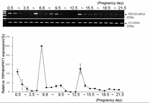
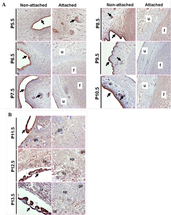
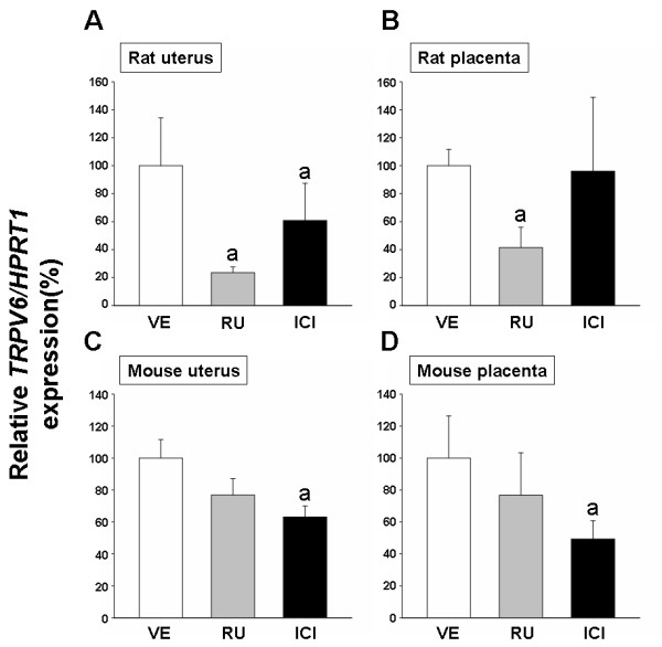
Transient receptor potential cation channel, subfamily V, member 6 (TRPV6) is an epithelial Ca2+ channel protein expressed in calcium absorbing organs. In the present study, we investigated the expression and regulation of uterine and placental TRPV6 during gestation in rodents. Uterine TRPV6 peaked at pregnancy day (P) 0.5, P5.5 and, P13.5 and was detected in uterine epithelium and glands of rats, while placental TRPV6 mRNA levels increased in mid-gestation. Uterine and placental TRPV6 mRNA levels in rats appear to cyclically change during pregnancy, suggesting that TRPV6 may participate in the implantation process. In addition, uterine TRPV6 mRNA is only expressed in placenta-unattached areas of the uterus, and uterine TRPV6 immunoreactivity was observed in luminal and glandular epithelial cells. In the placenta, TRPV6 was detected in the labyrinth and spongy zone. These results may indicate that TRPV6 has at least two functions: implantation of the embryo and maintenance of pregnancy. To investigate the pathway(s) mediating TRPV6 expression in rodents, anti-steroid hormone antagonists were injected prior to maximal TRPV6 expression. In rats, TRPV6 expression was reduced by RU486 (an anti-progesterone) through progesterone receptors, and ICI 182,780 (an anti-estrogen) blocked TRPV6 expression via estrogen receptors in mice. The juxtaposition of uterine and placental TRPV6 expressed in these tissues supports the notion that TRPV6 participates in transferring calcium ions between the maternal and fetal compartments. Taken together, TRPV6 gene may function as a key element in controlling calcium transport in the uterus between the embryo and the placenta during pregnancy.


Figures

Figure 1
Figure 1
Expression of uterine TRPV6 mRNA during pregnancy in rats. Uteri of gestating rats (n = 2) were collected daily from P0.5 to P21.5, and expression of TRPV6 mRNA was assayed by RT-PCR (Top panel, agarose gel image) and Real-Time PCR (Bottom panel, line graph). The unattached uteri containing uterine epithelial cells were used as a RNA preparation from P1.3.5. The line graph shows the analysis of Real-Time PCR data expressed as a percentage of TRPV6/HPRT1 (mean ± SEM of duplicates).
Figure 2
Figure 2
Placental TRPV6 mRNA expression during pregnancy in rats. Placentas were collected daily from P13.5 to P21.5. Placenta TRPV6 mRNA was examined by RT-PCR (Top panel, agarose gel image) and Real-Time PCR (Bottom panel, line graph). The line graph represents the analysis of Real-Time PCR data expressed as a percentage of TRPV6/HPRT1 (mean ± SEM of duplicates).
Figure 3
Figure 3
Localization of uterine TRPV6 at P6.5. Left panel, low power image of uterus showing representative TRPV6 immuno-positive regions restricted to three histological areas (A – C) as spotted rectangles. A, glandular epithelium; B, putative implantation site; C, epithelial layer. 'No first antibody' indicates immunoreactivity without anti-TRPV6 treatment, as a negative control. Arrows indicate immuno-positive staining.
Figure 4
Figure 4
Localization of placental TRPV6 at P20.5. Left panel, overview of placenta divided into three distinct areas (A to C) showing TRPV6 immuno-staining as spotted rectangles. A, inner labyrinth area; B, middle labyrinth; C, spongy zone. 'No first antibody' indicates immunoreactivity without anti-TRPV6 treatment, as a negative control. Arrows indicate immuno-positive staining.
Figure 5
Figure 5
Spatial expression of uterine (A) and placental (B) TRPV6 expression in a time-dependent manner. A, Rat uteri from P5.5 to P10.5 were separately shown into the non-attached or attached uteri using anit-TRPV6 serum. u, attached uterus; f, placenta-like structure; arrows, epithelial and glandular cells. B, Rat placentas from P11.5 to P13.5 were presented in a time-dependent manner. la, labyrinth zone; sp, spongy zone; gc, giant cells; arrows, fetal membrane.
Figure 6
Figure 6
Effects of steroid receptor antagonists on uterine and placental TRPV6 mRNA expressions. Panel A (uteri at P5.5) and B (placenta at P20.5) presented the rat TRPV6 mRNA levels. Four groups of pregnant rats (n = 4 per group) were treated with ethanol as a negative control (VE), progesterone receptor antagonist (RU, 2.5 mg per rat), or estrogen receptor antagonist (ICI, 0.5 mg per mouse). Panel C (uteri at P10.5) and D (placenta at P10.5) showed the mouse TRPV6 mRNA expressions. Four groups of pregnant mice (n = 4 per group) were treated with ethanol as a negative control (VE), progesterone receptor antagonist (RU, 25 μg per mouse) or estrogen receptor antagonist (ICI, 2 μg per mouse). Murine TRPV6 mRNA levels were examined by real-time PCR. The bar graph represents the analysis of real-time PCR data expressed as a percentage of TRPV6/HPRT1 (mean ± SEM of duplicates). a, statistically significant compared to a vehicle (P < 0.05).

References

    1. Wang J, Mayernik L, Armant DR. Integrin signaling regulates blastocyst adhesion to fibronectin at implantation: intracellular calcium transients and vesicle trafficking in primary trophoblast cells. Developmental biology. 2002;245:270–279. doi: 10.1006/dbio.2002.0644. - DOI - PubMed
    1. Wang J, Mayernik L, Armant DR. Trophoblast adhesion of the peri-implantation mouse blastocyst is regulated by integrin signaling that targets phospholipase C. Developmental biology. 2007;302:143–153. doi: 10.1016/j.ydbio.2006.09.015. - DOI - PMC - PubMed
    1. Hoenderop JG, Nilius B, Bindels RJ. Epithelial calcium channels: from identification to function and regulation. Pflugers Arch. 2003;446:304–308. - PubMed
    1. van Abel M, Huybers S, Hoenderop JG, Kemp AW van der, van Leeuwen JP, Bindels RJ. Age-dependent alterations in Ca2+ homeostasis: role of TRPV5 and TRPV6. Am J Physiol Renal Physiol. 2006;291:F1177–1183. doi: 10.1152/ajprenal.00038.2006. - DOI - PubMed
    1. Van Cromphaut SJ, Rummens K, Stockmans I, Van Herck E, Dijcks FA, Ederveen AG, Carmeliet P, Verhaeghe J, Bouillon R, Carmeliet G. Intestinal calcium transporter genes are upregulated by estrogens and the reproductive cycle through vitamin D receptor-independent mechanisms. J Bone Miner Res. 2003;18:1725–1736. doi: 10.1359/jbmr.2003.18.10.1725. - DOI - PubMed

Publication types

MeSH terms