

Reducing between scanner differences in multi-center PET studies
- PMID: 19457369
- PMCID: PMC4308413
- DOI: 10.1016/j.neuroimage.2009.01.057
Reducing between scanner differences in multi-center PET studies
Abstract
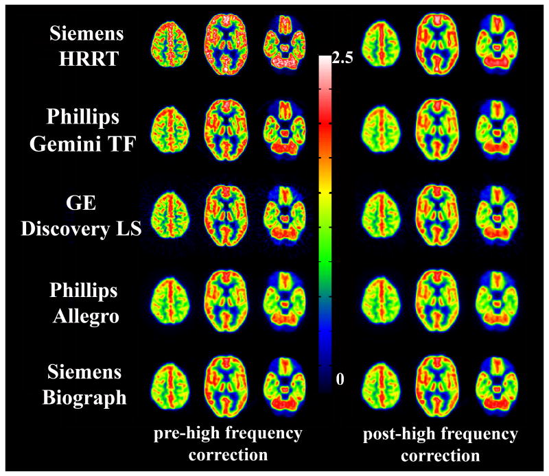
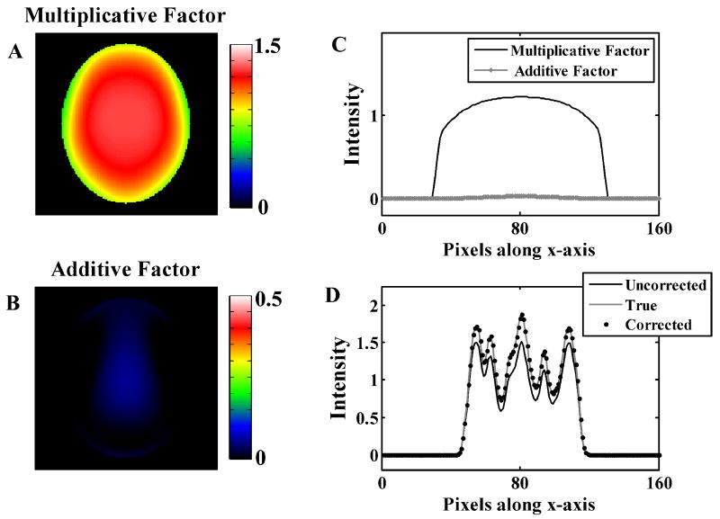
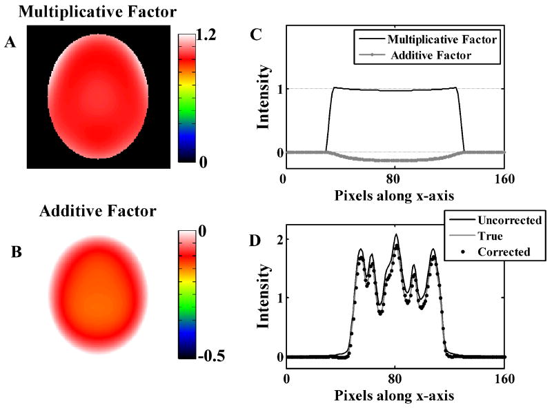
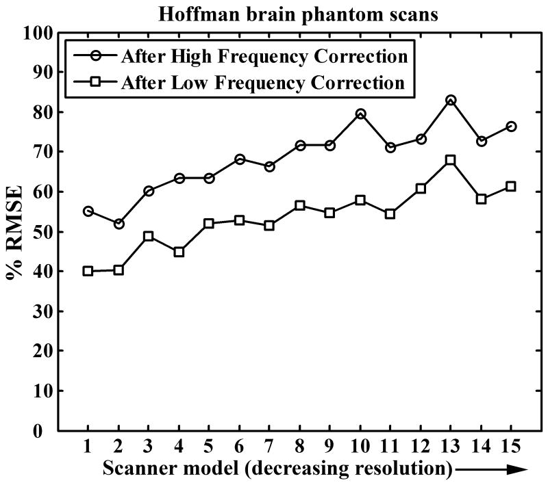
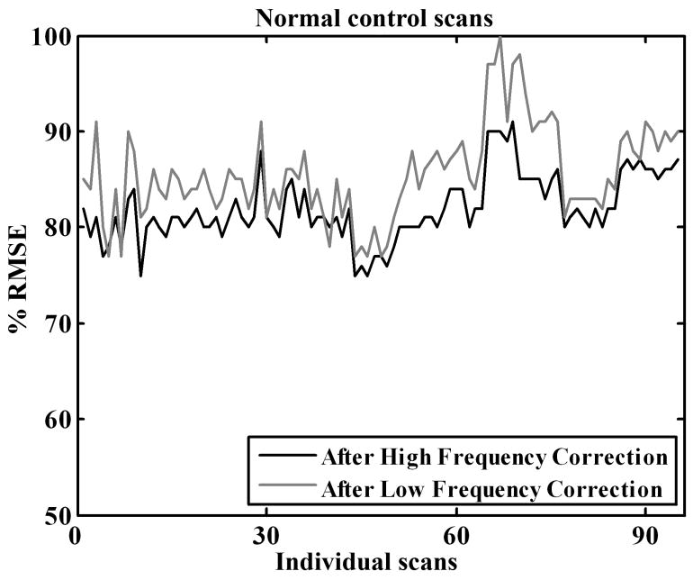
This work is part of the multi-center Alzheimer's Disease Neuroimaging Initiative (ADNI), a large multi-site study of dementia, including patients having mild cognitive impairment (MCI), probable Alzheimer's disease (AD), as well as healthy elderly controls. A major portion of ADNI involves the use of [(18)F]-fluorodeoxyglucose (FDG) with positron emission tomography (PET). The objective of this paper is the reduction of inter-scanner differences in the FDG-PET scans obtained from the 50 participating PET centers having fifteen different scanner models. In spite of a standardized imaging protocol, systematic inter-scanner variability in PET images from various sites is observed primarily due to differences in scanner resolution, reconstruction techniques, and different implementations of scatter and attenuation corrections. Two correction steps were developed by comparison of 3-D Hoffman brain phantom scans with the 'gold standard' digital 3-D Hoffman brain phantom: i) high frequency correction; where a smoothing kernel for each scanner model was estimated to smooth all images to a common resolution and ii) low frequency correction; where smooth affine correction factors were obtained to reduce the attenuation and scatter correction errors. For the phantom data, the high frequency correction reduced the variability by 20%-50% and the low frequency correction further reduced the differences by another 20%-25%. Correction factors obtained from phantom studies were applied to 95 scans from normal control subjects obtained from the participating sites. The high frequency correction reduced differences similar to the phantom studies. However, the low frequency correction did not further reduce differences; hence further refinement of the procedure is necessary.
Figures
References
-
- Fessler JA. Technical Report 293. Comm. and Sign. Proc. Lab., Dept. of EECS, Univ. of Michigan; Ann Arbor, MI: 1995. ASPIRE 3.0 user's guide: A sparse iterative reconstruction library; pp. 48109–2122.
-
- Hoffman EJ, Cutler PD, Digby WM, Mazziotta JC. 3-D phantom to simulate cerebral blood flow and metabolic images for PET. IEEE Trans Nucl Sci. 1990;37:616–620.
-
- Mazziotta JC, Toga AW, Evans A, Fox P, Lancaster J. A probabilistic atlas of the human brain: theory and rationale for its development. The International Consortium for Brain Mapping (ICBM) Neuroimage. 1995;2:89–101. - PubMed
-
- Minoshima S, Koeppe RA, Frey KA, Ishihara M, Kuhl DE. Stereotactic PET atlas of the human brain: aid for visual interpretation of functional brain images. J Nucl Med. 1994a;35:949–954. - PubMed
Publication types
MeSH terms
Substances
Grants and funding
LinkOut - more resources
Full Text Sources
Other Literature Sources
Medical
