. 2009 Jul 31;284(31):20869-75.
doi: 10.1074/jbc.M109.002923. Epub 2009 May 20.

Interferon-lambda is functionally an interferon but structurally related to the interleukin-10 family

Affiliations

Interferon-lambda is functionally an interferon but structurally related to the interleukin-10 family

Hans Henrik Gad et al. J Biol Chem. .

Abstract

Interferon-lambda (IFN-lambda) is an antiviral cytokine that signals through a distinct receptor complex, composed of the IFN-lambdaR1 and interleukin-10R2 (IL-10R2) receptor chains. We have determined the crystal structure of human IFN-lambda3 and characterized the interaction with its receptor complex through structure-based site-directed mutagenesis. The ability of IFN-lambda3 mutants to signal was determined by measuring the antiviral activity and induced STAT2 phosphorylation. In conclusion, our data show that, although IFN-lambda is functionally an interferon, it is clearly structurally related to members of the IL-10 family. In particular, we found an interesting similarity between IFN-lambda and IL-22, and we suggest that IFN-lambda and IL-22 possess parallel functions, protecting epithelial tissue against viral and bacterial infections, respectively.


Figures
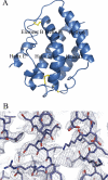
FIGURE 1.
FIGURE 1.
The crystal structure of human IFN-λ3. Upper panel, front view of the final model of IFN-λ3. Disulfide bonds are shown as sticks in yellow. Lower panel, electron density (final σA-weighted 2FoFc) for the molecule contoured at 1.0 σ and presented as blue mesh. Two iodide ions are shown in magenta.
FIGURE 2.
FIGURE 2.
Comparison of structures of class II cytokines. A, superimposition of IFN-α2 onto IFN-λ3. Ribbons of IFN-α and IFN-λ3 are colored green and blue, respectively. The two molecules have an r.m.s.d. of 4.1 Å. B, superimposition of IL-10 onto IFN-λ3. Ribbons of IL-10 and IFN-λ3 are colored magenta and blue, respectively. The two molecules have an r.m.s.d. of 2.9 Å. C, superimposition of IL-22 onto IFN-λ3. Ribbons of IL-22 and IFN-λ3 are colored brown and blue, respectively. The two molecules have an r.m.s.d. of 2.7 Å. Disulfide bonds are shown as sticks in yellow.
FIGURE 3.
FIGURE 3.
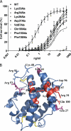
Characterization of the receptor binding site of IFN-λ3. A, antiviral activity of wild-type IFN-λ3 and selected mutants in HepG2 cells challenged with EMCV. Only mutants with EC50 values increasing >25-fold as compared with wild-type IFN-λ3 are shown. HepG2 cells were incubated with serial dilutions of IFN-λ3 before being challenged with EMCV. The data shown for wild-type IFN-λ3 represent four separate plates with each concentration done in replicas of four. Data shown for the mutants represent one plate with each concentration done in replicas of four. Mean ± S.D. from four replicates are shown. B, location of the mutated residues on the structure of IFN-λ3. Residues, which have been mutated to alanine, are shown as sticks. Mutations with EC50 values increasing <5-fold are shown in blue, whereas mutations with EC50 values increasing between 5- and 25-fold are shown in magenta, and, finally, mutations with EC50 values increasing >25-fold are shown in red.
FIGURE 4.
FIGURE 4.
STAT2 phosphorylation induced by wild-type IFN-λ3 and selected mutants in HepG2 cells correlates with antiviral activity. a, antiviral activity of wild-type IFN-λ3 in HepG2 cells challenged with EMCV correlates with STAT2 phosphorylation. The data shown for antiviral activity corresponds to the data shown in Fig. 3A. STAT2 phosphorylation was measured using the Luminex method at four different concentrations of IFN-λ3 corresponding to the EC25, EC50, EC75, and EC95. Mean ± S.D. from three replicates are shown. b, STAT2 phosphorylation induced by wild-type IFN-λ3 and selected mutants at EC80 concentrations in HepG2 cells. Mean ± S.D. from three replicates are shown.
FIGURE 5.
FIGURE 5.
Comparison of the receptor binding sites on IFN-λ3, IL-22, and IL-10. A, the receptor binding site of IL-10. Residues involved in binding to IL-10R1 and IL-10R2 as determined by crystallography and surface plasmon resonance, are shown in green and blue, respectively. The approximately location of the binding sites 1A and 1B are shown by circles. B, the receptor binding site of IL-22. Residues involved in binding to IL-22R1 and IL-10R2 as determined by crystallography and surface plasmon resonance, are shown in green and blue, respectively. The approximately location of the binding sites 1A and 1B are shown by circles. C, the receptor binding site of IFN-λ3. Residues involved in binding to its receptor, as determined by mutagenesis and antiviral activity, are shown in red. All molecules are shown from the same angle.

References

    1. Kotenko S. V., Gallagher G., Baurin V. V., Lewis-Antes A., Shen M., Shah N. K., Langer J. A., Sheikh F., Dickensheets H., Donnelly R. P. (2003) Nat. Immunol. 4, 69–77 - PubMed
    1. Sheppard P., Kindsvogel W., Xu W., Henderson K., Schlutsmeyer S., Whitmore T. E., Kuestner R., Garrigues U., Birks C., Roraback J., Ostrander C., Dong D., Shin J., Presnell S., Fox B., Haldeman B., Cooper E., Taft D., Gilbert T., Grant F. J., Tackett M., Krivan W., McKnight G., Clegg C., Foster D., Klucher K. M. (2003) Nat. Immunol. 4, 63–68 - PubMed
    1. Meager A., Visvalingam K., Dilger P., Bryan D., Wadhwa M. (2005) Cytokine 31, 109–118 - PubMed
    1. Robek M. D., Boyd B. S., Chisari F. V. (2005) J. Virol. 79, 3851–3854 - PMC - PubMed
    1. Ank N., West H., Bartholdy C., Eriksson K., Thomsen A. R., Paludan S. R. (2006) J. Virol. 80, 4501–4509 - PMC - PubMed

Publication types

LinkOut - more resources