. 2009 Jul 10;284(28):18882-92.
doi: 10.1074/jbc.M109.009886. Epub 2009 May 19.

Induction of the cytoprotective enzyme heme oxygenase-1 by statins is enhanced in vascular endothelium exposed to laminar shear stress and impaired by disturbed flow

Affiliations

Induction of the cytoprotective enzyme heme oxygenase-1 by statins is enhanced in vascular endothelium exposed to laminar shear stress and impaired by disturbed flow

Faisal Ali et al. J Biol Chem. .

Abstract

In addition to cholesterol-lowering properties, statins exhibit lipid-independent immunomodulatory, anti-inflammatory actions. However, high concentrations are typically required to induce these effects in vitro, raising questions concerning therapeutic relevance. We present evidence that endothelial cell sensitivity to statins depends upon shear stress. Using heme oxygenase-1 expression as a model, we demonstrate differential heme oxygenase-1 induction by atorvastatin in atheroresistant compared with atheroprone sites of the murine aorta. In vitro, exposure of human endothelial cells to laminar shear stress significantly reduced the statin concentration required to induce heme oxygenase-1 and protect against H(2)O(2)-mediated injury. Synergy was observed between laminar shear stress and atorvastatin, resulting in optimal expression of heme oxygenase-1 and resistance to oxidative stress, a response inhibited by heme oxygenase-1 small interfering RNA. Moreover, treatment of laminar shear stress-exposed endothelial cells resulted in a significant fall in intracellular cholesterol. Mechanistically, synergy required Akt phosphorylation, activation of Kruppel-like factor 2, NF-E2-related factor-2 (Nrf2), increased nitric-oxide synthase activity, and enhanced HO-1 mRNA stability. In contrast, heme oxygenase-1 induction by atorvastatin in endothelial cells exposed to oscillatory flow was markedly attenuated. We have identified a novel relationship between laminar shear stress and statins, demonstrating that atorvastatin-mediated heme oxygenase-1-dependent antioxidant effects are laminar shear stress-dependent, proving the principle that biomechanical signaling contributes significantly to endothelial responsiveness to pharmacological agents. Our findings suggest statin pleiotropy may be suboptimal at disturbed flow atherosusceptible sites, emphasizing the need for more specific therapeutic agents, such as those targeting Kruppel-like factor 2 or Nrf2.


Figures
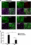
FIGURE 1.
FIGURE 1.
Atorvastatin induces endothelial HO-1 expression in murine aortic EC. HO-1 expression in murine aortic endothelium of C57BL/6 mice assessed en face using anti-HO-1 (red). EC were identified by fluorescein isothiocyanate-conjugated anti-CD31 (green) and cell nuclei with Draq5 (purple). An isotype-matched control antibody did not bind (not shown). A and B, representative images of HO-1 staining from: A, the greater curvature (low probability (LP) area); and B, the lesser curvature (high probability (HP) area). C, HO-1 expression was quantified (mean ± S.D.) by image analysis of fluorescence intensity in multiple cells at 3 distinct sites and expressed as EC fluorescence above a threshold intensity defined by background fluorescence. *, p < 0.05.
FIGURE 2.
FIGURE 2.
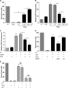
LSS and statins synergistically enhance HO-1 expression. HUVEC were exposed to static conditions (gray bars) or unidirectional LSS (12 dynes/cm2) (black bars) for 24 h. After 12 h, statin or vehicle was added to the culture medium. A, atorvastatin (AT) 2.5 μm; B, AT 0.6 μm; dotted lines represent the predicted HO-1 mRNA level achieved by an additive response between LSS and atorvastatin. C, dose-response for AT, # = synergistic response, § = additive response, with HO-1 RNA quantified by real-time PCR. Dotted line represents HO-1 mRNA induced by LSS alone. D, HUVEC were transfected with the pHO-1 luciferase reporter construct or pGL3-basic, prior to exposure to static conditions or unidirectional LSS and addition of AT 0.6 μm or vehicle as above and analysis of luciferase activity at 24 h. E, HUVEC were exposed to static culture or LSS for 24 h with atorvastatin added after 12 h and HO-1 analyzed by immunoblotting. F, HUVEC were exposed to static conditions (gray bars), LSS (12 dynes/cm2) (black bars), or OF (1 Hz, ±5 dynes/cm2) (hatched bars) for 24 h. After 12 h, statin or vehicle was added to the culture medium. Data are expressed as mean ± S.E. from three experiments. *, p < 0.05; **, p < 0.01. UT, untreated.
FIGURE 3.
FIGURE 3.
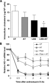
LSS and statins synergistically enhance resistance to oxidative stress. HUVEC were exposed to static conditions (gray bars) or LSS (12 dynes/cm2) (black bars) for 24 h. After 12 h, statin or vehicle was added to the culture medium. A, HUVEC were treated with H2O2 (50 μm) or vehicle for 45 min. Live cells were quantified by trypan blue exclusion, expressed as a percentage of untreated (UT) control (n = 3). B and C, cells were treated as in A, followed by loading with CM-H2DCFDA (5 μm) and exposure to: B, H2O2 (5 μm) for 30 min; or C, leptin (100 ng/ml) for 120 min. Oxidative stress was quantified by flow cytometric analysis (n = 3). D, HUVEC were left untreated or transfected with scrambled siRNA (CT) or HO-1 siRNA for 24 h, prior to exposure to LSS and atorvastatin (0.6 μm) for 24 h as in A. EC were treated with H2O2 (50 μm) as above and live cells were quantified by MTT assay (percent untreated control, n = 4). E, HUVEC were exposed to static conditions (gray bars) or OF (1 Hz, ±5 dynes/cm2) (hatched bars) for 24 h. After 12 h, atorvastatin (0.6 μm) (AT) or vehicle were added to the culture medium. After exposure to H2O2 live cells were quantified by MTT assay. Data are expressed as mean ± S.E. from three experiments. *, p < 0.05; **, p < 0.01.
FIGURE 4.
FIGURE 4.
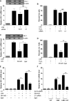
Statin and LSS reduce cholesterol and enhance HO-1 mRNA stability. HUVEC were exposed to static conditions or LSS (12 dynes/cm2) for 24 h. After 12 h, atorvastatin (AT) (0.6 μm) or vehicle were added to the culture medium. A, EC were homogenized and cholesterol content was analyzed by mass spectrometry and expressed as μg/106 cells. B, HUVEC monolayers were divided into sections using a water-resistant pen and actinomycin D (2 μg/ml) or vehicle added. EC were harvested after 0–6 h for RNA extraction. HO-1 mRNA was quantified by quantitative RT-PCR, plotted as a percentage of mRNA expression prior to the addition of actinomycin D. Data are presented as mean ± S.E. (n = 3). *, p < 0.05. UT, untreated.
FIGURE 5.
FIGURE 5.
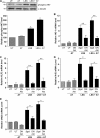
Synergistic HO-1 induction by LSS requires KLF2 and Nrf2. A and B, HUVEC were left untransfected (UT) or transfected with scrambled siRNA (CT) or KLF2 siRNA, prior to exposure to static conditions or LSS (12 dynes/cm2) for 24 h. After 12 h, atorvastatin (0.6 μm) or vehicle were added to the culture medium. A, HO-1 mRNA was quantified by real-time PCR, a representative cDNA gel of PCR products is shown. B, following exposure to atorvastatin and LSS, HUVEC were treated with H2O2 (50 μm) or vehicle for 45 min. Live cells were quantified by MTT assay (percent untreated control, n = 4). C and D, HUVEC were left untransfected (UT) or transfected with an adenovirus expressing β-galactosidase (βgal) or DN-Nrf2 prior to exposure to static conditions or LSS (12 dynes/cm2) for 24 h. C, HO-1 mRNA was quantified by real-time PCR. D, HUVEC were treated with H2O2 (50 μm) or vehicle for 45 min and live cells were quantified by MTT assay. E and F, HUVEC were left untransfected or transfected with: E, β-galactosidase or DN-Nrf2 adenovirus; or F, control siRNA (CT), KLF2 siRNA, β-galactosidase control, or DN-Nrf2 adenovirus prior to exposure to static conditions (gray bars) or LSS (black bars) for 24 h with addition of vehicle or atorvastatin (AT) (0.6 μm) after 12 h. HO-1 mRNA was quantified by real-time PCR. Data are expressed as mean ± S.E. from three experiments. *, p < 0.05; **, p < 0.01.
FIGURE 6.
FIGURE 6.
Synergistic induction of HO-1 is dependent upon Akt. HUVEC were exposed to static conditions (gray bars) or unidirectional LSS (12 dynes/cm2) (black bars) for 24 h. After 12 h, atorvastatin (0.6 μm) (AT) or vehicle control was added. A, EC were immunoblotted with antibodies against phosphorylated-Akt (Ser473) and α-tubulin. The histogram shows phospho-Akt expression quantified by densitometry relative to the α-tubulin bands. B–E, HUVEC were left untransfected (UT) or transfected with a β-galactosidase control adenovirus (βgal) or DN-Akt adenovirus prior to exposure to static conditions or LSS (12 dynes/cm2) for 24 h as above. HO-1 (B), KLF2 (C), Nrf2 (D), and eNOS (E) mRNA levels were quantified by real-time PCR. Data are expressed as mean ± S.E. from three experiments. *, p < 0.05; **, p < 0.01.
FIGURE 7.
FIGURE 7.
Synergistic induction of HO-1 is dependent upon activity of eNOS. HUVEC were left untreated (UT) or pre-treated with l-NAME (L-N) (1 mm) or vehicle prior to exposure to static conditions or unidirectional LSS (12 dynes/cm2) for 24 h with atorvastatin (0.6 μm) (AT) or vehicle control added after 12 h. A, HO-1; and B, thrombomodulin (TM) mRNA levels were analyzed by real-time PCR; or C, Nrf2 activation was assessed in EC nuclear extracts by analysis of Nrf2 binding to the antioxidant responsive element, quantified using a TransAM assay. Data are expressed as mean ± S.E. from two to three experiments. *, p < 0.05; **, p < 0.01.

References

    1. Jain M. K., Ridker P. M. (2005) Nat. Rev. Drug Discov. 4, 977–987 - PubMed
    1. Landmesser U., Bahlmann F., Mueller M., Spiekermann S., Kirchhoff N., Schulz S., Manes C., Fischer D., de Groot K., Fliser D., Fauler G., März W., Drexler H. (2005) Circulation 111, 2356–2363 - PubMed
    1. Ridker P. M., Danielson E., Fonseca F. A., Genest J., Gotto A. M., Jr., Kastelein J. J., Koenig W., Libby P., Lorenzatti A. J., MacFadyen J. G., Nordestgaard B. G., Shepherd J., Willerson J. T., Glynn R. J. (2008) N. Engl. J. Med. 359, 2195–2207 - PMC - PubMed
    1. Mason J. C. (2003) Clin. Sci. 105, 251–266 - PubMed
    1. Liao J. K., Laufs U. (2005) Annu. Rev. Pharmacol. Toxicol. 45, 89–118 - PMC - PubMed

Publication types

MeSH terms