. 2009 Aug 1;83(3):481-92.
doi: 10.1093/cvr/cvp148. Epub 2009 May 20.

Targeted G-protein inhibition as a novel approach to decrease vagal atrial fibrillation by selective parasympathetic attenuation

Affiliations

Targeted G-protein inhibition as a novel approach to decrease vagal atrial fibrillation by selective parasympathetic attenuation

Gary L Aistrup et al. Cardiovasc Res. .

Abstract

Aims: The parasympathetic nervous system is thought to play a key role in atrial fibrillation (AF). Since parasympathetic signalling is primarily mediated by the heterotrimeric G-protein, Galpha(i)betagamma, we hypothesized that targeted inhibition of Galpha(i) interactions in the posterior left atrium (PLA) would modify the substrate for vagal AF.

Methods and results: Cell-penetrating(cp)-Galpha(i)1/2 and cp-Galpha(i)3 C-terminal peptides were assessed for their ability to attenuate cholinergic-parasympathetic signalling in isolated feline atrial myocytes and in canine left atrium (LA). Confocal fluorescence microscopy indicated that cp-Galpha(i)1/2 and/or cp-Galpha(i)3 peptides moderated carbachol attenuation of cellular Ca(2+) transients in isolated atrial myocytes. High-density epicardial mapping of dog PLA, left atrial pulmonary veins (PVs), and left atrial appendage (LAA) indicated that the delivery of cp-Galpha(i)1/2 peptide or cp-Galpha(i)3 peptide into the PLA prolonged effective refractory periods at baseline and during vagal stimulation in the PLA and to varying extents also in the LAA and PV regions. After delivery of cp-Galpha(i) peptides into the PLA, AF inducibility during vagal stimulation was significantly diminished.

Conclusion: These results demonstrate the feasibility of using specific G(i)-protein inhibition to achieve selective parasympathetic denervation in the PLA, with a resulting change in vagal responsiveness across the entire LA.


Figures
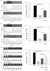
Figure 1
Figure 1
CCh attenuation of CaTs in atrial myocytes is blunted in the presence of cp-Gαi peptides. (A) Serial confocal X-t linescan images (fluo-4 fluorescence) and the corresponding control (no CCh) peak-normalized fluorescence (CPNF) vs. t profiles illustrating an example of an individual isolated feline myocyte paired responses to: (i) acute application of 10 µM CCh and subsequent washout-recovery in the absence of peptide; (ii) application of 10 µM CCh after pre-1 min and co-application of cp-Gαi1/2 peptide showing blunting of the CCh action (CCh was applied for ≤3 s to ensure effect was sustained); and (iii) summary statistics bar graph of the paired responses from 10 cells in terms of peptide effect on CaT peak amplitude. (B) As in (A), but using cp-Gαi3 peptide in a different myocyte (in ii). Summary statistics bar graph (in iii) compiled from nine cells. (C) As in (A), but in which a different myocyte was exposed first (in ii) to cp-Gαi1/2 peptide, and then (in iii) to cp-Gαi3 peptide. Summary statistics bar graph in (iv) compiled from four cells. NS, not significant. *P < 0.05, **P < 0.01, and ***P < 0.001.
Figure 2
Figure 2
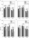
Effect of cp-Gαi peptides on left atrial ERPs. Summary statistics for ERPs at baseline (BL) and during VS in the PLA, PVs, and LAA are as follows: (A) ± cp-Gαi1/2 peptide delivery (sono/electroporation-assisted) into the PLA B) ± cp-Gαi3 peptide delivery (electroporation-assisted) into the PLA; C) ± cp-GαoR peptide delivery (electroporation-assisted) into the PLA; and D) ± sono- or electroporation of the PLA alone. NS, not significant. *P < 0.05, **P < 0.01, and ***P < 0.001.
Figure 3
Figure 3
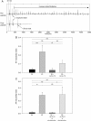
Effect of cp-Gαi peptide on AF inducibility. (A) Example electrogram recordings from the same epicardial electrode in the PLA in which the upper tracing shows that after delivery of a single premature stimulus (S2) during VS, induced an episode of AF in the absence of cp-Gαi1/2 peptide; whereas the lower tracing shows that in another episode of AF, this manoeuvre did not induce AF in the presence of cp-Gαi1/2 peptide delivered into the PLA. (B) Statistical summary of AF inducibility ratio at baseline (BL) and during VS in the left atrium ± cp-Gαi peptides delivery into the PLA (combinatorial average of cp-Gαi1/2 and cp-Gαi3 peptide results). (C) Statistical summary of AF inducibility ratio at baseline and during VS in left atrium ± cp-GαoR peptide delivery into the PLA or sono/electroporation of PLA (combinatorial average of results from these manoeuvres).
Figure 4
Figure 4
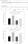
Effect of cp-Gαi peptide on the dominant frequency and organization of AF. (A) Examples of electrogram recordings from the same epicardial electrode in the PLA in which the upper tracing shows an episode of AF induced during VS in the absence of cp-Gαi1/2 peptide; whereas the lower tracing shows another episode of AF induced during VS after cp-Gαi1/2 peptide delivery into the PLA. Clearly, the most predominant frequency component, or dominant frequency (DF), of the AF episode in the presence of peptide is lower (has a slower periodicity) compared with that in the absence of peptide. (B) Statistical summary of the DF of AF in the absence of (AF−VS) and during VS (AF+VS) in the PLA, PV, and LAA regions ± cp-Gαi1/2 peptide delivery into the PLA. (C) Statistical summary of DF of AF in the absence of and during VS in left atrium ± cp-GαoR peptide delivery into the PLA or sono/electroporation of PLA (combinatorial average of results from these manoeuvres).
Figure 5
Figure 5
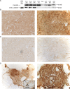
Retention of cp-Gαi1/2 peptide in the PLA of cpGPp dogs. (A) Example of anti-FLAG western blots of samples as specified (above each lane): ‘EP’, atrial samples taken from dogs subjected to in vivo electrophysiology experiments in which FLAG-tagged cp-Gαi1/2 peptide had been delivered into their PLAs; ‘–’, atrial samples taken from dog in which no peptides had been delivered—i.e. negative controls; the amount of pure FLAG-tagged cp-Gαi1/2 peptide used in lane 1 was 1 µg; ‘numbers’ to the right of PLA or LAA specifiers refer to the particular dogs. (B) (Left) 10X bright field image of a 5–10 µm thick cross-section (endocardium-to-epicardium) of the area of PLA that had been injected with 1 µM FLAG-tagged cp-Gαi1/2 during an in vivo electrophysiology experiment (see text) showing marked brown anti-FLAG secondary horseradish-peroxidase (HRP) staining throughout; and (right) a region within the latter at 40X myocardium. (C) As in (B), but of a section of LAA—region remote from the PLA site of peptide injection—showing essentially only background HRP staining. (D) As in (B), but of a section of PLA near the area of peptide delivery that contained an autonomic nerve bundle within a fat pad (left inset is 4X field at ⅓ scale); and the region within the latter at 40X showing ganglion cells within the nerve bundle. The marked brown anti-FLAG secondary HRP staining indicates that FLAG-tagged cp-Gαi1/2 peptide had incorporated into this nerve bundle. Light green staining (methyl-green) in these sections denotes staining of cell nuclei. Photomicrographs shown were taken at identical camera settings/exposures.
Figure 6
Figure 6
cAMP in cpGPp dogs vs. control dogs. Statistical summary of cAMP in PLA and LAA tissue explant homogenates from dogs whose PLAs had been injected (+electroporation) with cp-Gαi1/2 peptide (cpGPp) compared with dogs whose PLAs were not injected with peptide (controls) during the in vivo electrophysiology experiments. LAAs were assayed as internal controls, particularly for cpGPp dogs.

References

    1. Conway E, Musco S, Kowey PR. New horizons in antiarrhythmic therapy: will novel agents overcome current deficits? Am J Cardiol. 2008;102:12H–19H. - PubMed
    1. Jais P, Cauchemez B, Macle L, Daoud E, Khairy P, Subbiah R, et al. Catheter ablation versus antiarrhythmic drugs for atrial fibrillation: the A4 study. Circulation. 2008;118:2498–2505. - PubMed
    1. Pokushalov E. The role of autonomic denervation during catheter ablation of atrial fibrillation. Curr Opin Cardiol. 2008;23:55–59. - PubMed
    1. Chou C-C, Chen P-S. New concepts in atrial fibrillation: mechanism and remodeling. Med Clin North Am. 2008;92:53–63. - PMC - PubMed
    1. Arora R, Ng J, Ulphani J, Mylonas I, Subacius H, Shade G, et al. Unique autonomic profile of the pulmonary veins and posterior left atrium. J Am Coll Cardiol. 2007;49:1340–1348. - PubMed

Publication types

MeSH terms