

Formation of syncytia is repressed by tetraspanins in human immunodeficiency virus type 1-producing cells
- PMID: 19458002
- PMCID: PMC2708618
- DOI: 10.1128/JVI.00163-09
Formation of syncytia is repressed by tetraspanins in human immunodeficiency virus type 1-producing cells
Abstract
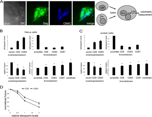
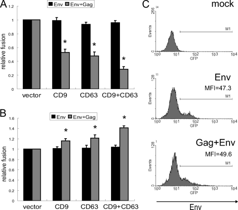
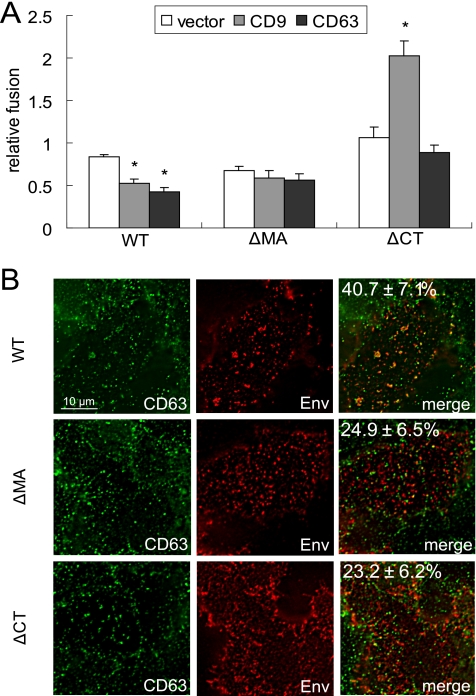
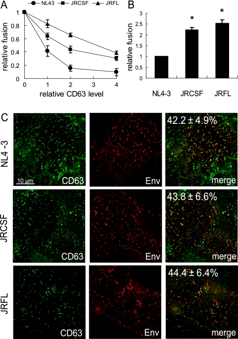
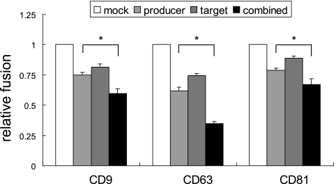
In vitro propagation studies have established that human immunodeficiency virus type 1 (HIV-1) is most efficiently transmitted at the virological synapse that forms between producer and target cells. Despite the presence of the viral envelope glycoprotein (Env) and CD4 and chemokine receptors at the respective surfaces, producer and target cells usually do not fuse with each other but disengage after the viral particles have been delivered, consistent with the idea that syncytia, at least in vitro, are not required for HIV-1 spread. Here, we tested whether tetraspanins, which are well known regulators of cellular membrane fusion processes that are enriched at HIV-1 exit sites, regulate syncytium formation. We found that overexpression of tetraspanins in producer cells leads to reduced syncytium formation, while downregulation has the opposite effect. Further, we document that repression of Env-induced cell-cell fusion by tetraspanins depends on the presence of viral Gag, and we demonstrate that fusion repression requires the recruitment of Env by Gag to tetraspanin-enriched microdomains (TEMs). However, sensitivity to fusion repression by tetraspanins varied for different viral strains, despite comparable recruitment of their Envs to TEMs. Overall, these data establish tetraspanins as negative regulators of HIV-1-induced cell-cell fusion, and they start delineating the requirements for this regulation.
Figures
Similar articles
-
Human immunodeficiency virus type 1 assembly, budding, and cell-cell spread in T cells take place in tetraspanin-enriched plasma membrane domains.J Virol. 2007 Aug;81(15):7873-84. doi: 10.1128/JVI.01845-06. Epub 2007 May 23. J Virol. 2007. PMID: 17522207 Free PMC article.
-
EWI-2 Inhibits Cell-Cell Fusion at the HIV-1 Virological Presynapse.Viruses. 2019 Nov 20;11(12):1082. doi: 10.3390/v11121082. Viruses. 2019. PMID: 31757023 Free PMC article.
-
Human immunodeficiency virus type 1 and influenza virus exit via different membrane microdomains.J Virol. 2007 Nov;81(22):12630-40. doi: 10.1128/JVI.01255-07. Epub 2007 Sep 12. J Virol. 2007. PMID: 17855546 Free PMC article.
-
The roles of tetraspanins in HIV-1 replication.Curr Top Microbiol Immunol. 2009;339:85-102. doi: 10.1007/978-3-642-02175-6_5. Curr Top Microbiol Immunol. 2009. PMID: 20012525 Free PMC article. Review.
-
Tetraspanins, Another Piece in the HIV-1 Replication Puzzle.Front Immunol. 2018 Aug 3;9:1811. doi: 10.3389/fimmu.2018.01811. eCollection 2018. Front Immunol. 2018. PMID: 30127789 Free PMC article. Review.
Cited by
-
Tetraspanins: Host Factors in Viral Infections.Int J Mol Sci. 2021 Oct 27;22(21):11609. doi: 10.3390/ijms222111609. Int J Mol Sci. 2021. PMID: 34769038 Free PMC article. Review.
-
Relationships between plasma membrane microdomains and HIV-1 assembly.Biol Cell. 2010 Mar 25;102(6):335-50. doi: 10.1042/BC20090165. Biol Cell. 2010. PMID: 20356318 Free PMC article. Review.
-
Macrophages and Cell-Cell Spread of HIV-1.Viruses. 2010 Aug 1;2(8):1603-1620. doi: 10.3390/v2081603. Viruses. 2010. PMID: 21552427 Free PMC article.
-
Comparative proteomic analysis of HIV-1 particles reveals a role for Ezrin and EHD4 in the Nef-dependent increase of virus infectivity.J Virol. 2013 Apr;87(7):3729-40. doi: 10.1128/JVI.02477-12. Epub 2013 Jan 16. J Virol. 2013. PMID: 23325686 Free PMC article.
-
HIV-1 Cell-Free and Cell-to-Cell Infections Are Differentially Regulated by Distinct Determinants in the Env gp41 Cytoplasmic Tail.J Virol. 2015 Sep;89(18):9324-37. doi: 10.1128/JVI.00655-15. Epub 2015 Jul 1. J Virol. 2015. PMID: 26136566 Free PMC article.
References
-
- Berditchevski, F., and E. Odintsova. 2007. Tetraspanins as regulators of protein trafficking. Traffic 889-96. - PubMed
-
- Bromley, S. K., W. R. Burack, K. G. Johnson, K. Somersalo, T. N. Sims, C. Sumen, M. M. Davis, A. S. Shaw, P. M. Allen, and M. L. Dustin. 2001. The immunological synapse. Annu. Rev. Immunol. 19375-396. - PubMed
Publication types
MeSH terms
Substances
Grants and funding
LinkOut - more resources
Full Text Sources
Medical
Research Materials
Miscellaneous
