. 2009 Aug;83(15):7375-83.
doi: 10.1128/JVI.00331-09. Epub 2009 May 20.

Cryo-electron microscopy structure of adenovirus type 2 temperature-sensitive mutant 1 reveals insight into the cell entry defect

Affiliations

Cryo-electron microscopy structure of adenovirus type 2 temperature-sensitive mutant 1 reveals insight into the cell entry defect

Mariena Silvestry et al. J Virol. 2009 Aug.

Abstract

The structure of the adenovirus type 2 temperature-sensitive mutant 1 (Ad2ts1) was determined to a resolution of 10 A by cryo-electron microscopy single-particle reconstruction. Ad2ts1 was prepared at a nonpermissive temperature and contains the precursor forms of the capsid proteins IIIa, VI, and VIII; the core proteins VII, X (mu), and terminal protein (TP); and the L1-52K protein. Cell entry studies have shown that although Ad2ts1 can bind the coxsackievirus and Ad receptor and undergo internalization via alphav integrins, this mutant does not escape from the early endosome and is targeted for degradation. Comparison of the Ad2ts1 structure to that of mature Ad indicates that Ad2ts1 has a different core architecture. The Ad2ts1 core is closely associated with the icosahedral capsid, a connection which may be mediated by preproteins IIIa and VI. Density within hexon cavities is assigned to preprotein VI, and membrane disruption assays show that hexon shields the lytic activity of both the mature and precursor forms of protein VI. The internal surface of the penton base in Ad2ts1 appears to be anchored to the core by interactions with preprotein IIIa. Our structural analyses suggest that these connections to the core inhibit the release of the vertex proteins and lead to the cell entry defect of Ad2ts1.


Figures
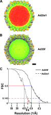
FIG. 1.
FIG. 1.
Cryo-EM reconstructions of Ad2ts1 and Ad35f reveal a major structural difference in the core of the virion. (A) Cropped view of the Ad2ts1 reconstruction. The crop plane is colored by the density value, with the strongest density in red and the weakest in green. The protein/DNA-containing core displays predominantly strong density (red). (B) Cropped view of the Ad35f reconstruction (38) with the crop plane colored as in panel A. Both reconstructions are shown filtered to 10.5-Å resolution. Scale bar, 100 Å. (C) An FSC plot indicating a resolution range for Ad2ts1 of 10.5 Å to 8.6 Å (10.5 Å at FSC 0.5, 9.5 Å at FSC 0.3, and 8.6 Å at FSC 0.143). The resolution range for Ad35f is 6.9 Å to 5.2 Å (6.9 Å at FSC 0.5, 6.1 Å at FSC 0.3, and 5.2 Å at FSC 0.143).
FIG. 2.
FIG. 2.
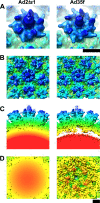
The main differences between Ad2ts1 and Ad35f are on the interior of the icosahedral capsid. (A) Views of the penton base with a segment of protruding fiber. The fiber shafts of both virions are flexible, and thus only short portions are reconstructed. Both structures are shown filtered to 10.5-Å resolution and radially color coded (300 Å = red; 480 Å = blue). (B) Outer views of the vertex regions showing a penton base with five surrounding hexons. (C) Side views of the two vertex regions at similar isosurface contour levels. The Ad2ts1 capsid (yellow to blue) is closely associated with and connects to the core of the virion (red). In contrast, the Ad35f capsid is separated from the core by a gap in the density. (D) Inner views of the vertex regions showing the strong core density for Ad2ts1 and the resolved internal capsid density below the penton base and surrounding hexons for Ad35f. Scale bars, 50 Å.
FIG. 3.
FIG. 3.
Average radial density distributions of the Ad2ts1 and Ad35f structures. Profiles for Ad2ts1 (solid line) and Ad35f (dashed line) were calculated with the IMAGIC-5 Threed-radial-density-options routine. The two profiles were normalized in the radial shell (370 to 463 Å) indicated by the bracket and corresponding to the outer portion of the icosahedral capsid.
FIG. 4.
FIG. 4.
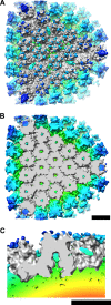
Preprotein VI is assigned to density within the cavity of every hexon trimer in Ad2ts1. (A) An enlarged facet (gray) consisting of docked crystal structures of 18 hexon trimers and 3 penton base pentamers is shown filtered to 10.5-Å resolution. The facet is superimposed on the Ad2ts1 difference density, which is radially color coded as in Fig. 2. The external difference density includes the protruding fiber shaft, surface loops of hexon and penton base missing from their respective crystal structures, and density assigned to protein IX. (B) Cropping away the top ∼80 Å of the facet reveals difference density inside of every hexon trimer (green). Similar density within the Ad35f hexons has been assigned to protein VI (38). The density within every hexon of Ad2ts1 is tentatively assigned as preprotein VI. (C) Side cropped view of a peripentonal hexon within the facet (gray) with the internal difference density in the shape of a “plug,” which connects to the Ad2ts1 core (yellow to red). Scale bars, 100 Å.
FIG. 5.
FIG. 5.
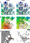
Density assigned to protein IX and preproteins IIIa and VIII is found within Ad2ts1. (A) The Ad2ts1 difference map (filtered to 10.5 Å) and the Ad35f difference map (filtered to 10.5 Å resolution) are shown superimposed on a portion of the facet (gray). The density assigned to the N-terminal domain of protein IX is outlined with triangles, and that assigned to the C-terminal domain of protein IX is within ovals (38). (B) Cropping away the top ∼100 Å reveals internal difference density below the penton base and hexons assigned to preproteins IIIa (oval) and VIII (rectangle) in Ad2ts1 and the mature forms of proteins IIIa and VIII in Ad35f. (C) Rotating by 180° and enlarging one copy of preprotein VIII (left, transparent) or mature protein VIII (right, transparent) shows the density rod assigned to the predicted 22-aa α-helix (red) in fragment 1 of protein VIII. In panel C the Ad35f density is shown filtered to 6.9 Å so that α-helical rod is well resolved. The internal Ad2ts1 difference density in panels B and C is shown with a relatively high isosurface contour level (0.65σ, versus 0.37σ in panel A) so that the density assigned to precursor proteins can be resolved from the core. The Ad35f difference density is shown with an isosurface contour level of 0.85σ to reveal the α-helical nature of the C-terminal domain of proteins IX, as well as proteins IIIa and VIII. In panels A and B the Ad2ts1 and Ad35f difference density maps are shown radially color coded as in Fig. 2. Scale bar, 50 Å.
FIG. 6.
FIG. 6.
Hexon binding shields protein VI and preprotein VI membrane lytic activities. Recombinant protein VI or preprotein VI was incubated with increasing amounts of purified hexon for 30 min before addition to SulfoB-containing liposomes at 37°C. The percentage of SulfoB released was measured 15 min after protein addition to liposomes. Protein VI (black circles) or preprotein VI (white circles) was present at a final concentration of 20 nM. Error bars represent standard errors of the means.
FIG. 7.
FIG. 7.
Diagram of proposed cell entry events for Ad2 and Ad5 versus Ad2ts1. (A) One vertex of an Ad2 or Ad5 virion is shown schematically (left). The location of protein IIIa (magenta ovals) underneath the penton base is as assigned by a cryo-EM study (38) and confirmed by a peptide tagging study (39). The position of protein VI (red) within the cavity of every hexon trimer is as assigned by cryo-EM (38). We propose that a critical step of disassembly is either release of the fiber or release of the fiber/penton base complex (middle). Vertex release is associated with release of 25% of the hexons and 80% of protein VI (right), as shown by Wiethoff et al. (50) for Ad5 at temperatures over 45°C. (B) One vertex of an Ad2ts1 virion is shown schematically, with preprotein IIIa (black ovals) and preprotein VI (orange) anchored to the immature DNA/protein core (cyan). The association of preprotein IIIa with both the N-terminal tails of penton base and the viral core may impede the release of the vertex proteins (fiber, penton base, and preprotein IIIa). In addition, anchoring of preprotein VI to the core may block its release.

References

    1. Adiga, U., W. T. Baxter, R. J. Hall, B. Rockel, B. K. Rath, J. Frank, and R. Glaeser. 2005. Particle picking by segmentation: a comparative study with SPIDER-based manual particle picking. J. Struct. Biol. 152211-220. - PubMed
    1. Anderson, C. W. 1990. The proteinase polypeptide of adenovirus serotype 2 virions. Virology 177259-272. - PubMed
    1. Bergelson, J. M., J. A. Cunningham, G. Droguett, E. A. Kurt-Jones, A. Krithivas, J. S. Hong, M. S. Horwitz, R. L. Crowell, and R. W. Finberg. 1997. Isolation of a common receptor for coxsackie B viruses and adenoviruses 2 and 5. Science 2751320-1323. - PubMed
    1. Brown, M. T., W. J. McGrath, D. L. Toledo, and W. F. Mangel. 1996. Different modes of inhibition of human adenovirus proteinase, probably a cysteine proteinase, by bovine pancreatic trypsin inhibitor. FEBS Lett. 388233-237. - PubMed
    1. Challberg, M. D., S. V. Desiderio, and T. J. Kelly, Jr. 1980. Adenovirus DNA replication in vitro: characterization of a protein covalently linked to nascent DNA strands. Proc. Natl. Acad. Sci. USA 775105-5109. - PMC - PubMed

Publication types

MeSH terms