. 2009 Aug;83(15):7397-410.
doi: 10.1128/JVI.00656-09. Epub 2009 May 20.

Utilization of immunoglobulin G Fc receptors by human immunodeficiency virus type 1: a specific role for antibodies against the membrane-proximal external region of gp41

Affiliations

Utilization of immunoglobulin G Fc receptors by human immunodeficiency virus type 1: a specific role for antibodies against the membrane-proximal external region of gp41

Lautaro G Perez et al. J Virol. 2009 Aug.

Abstract

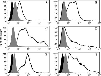
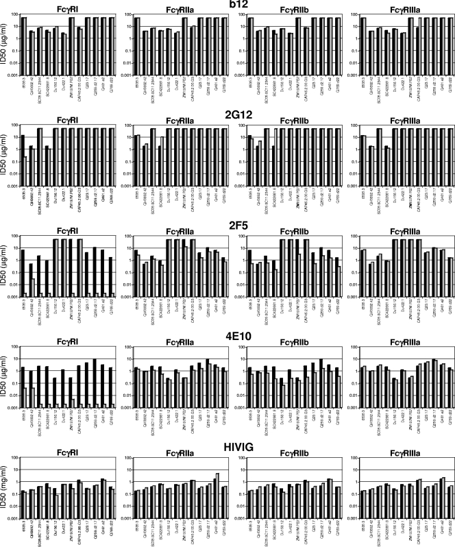
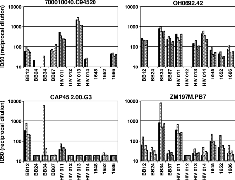
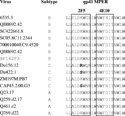
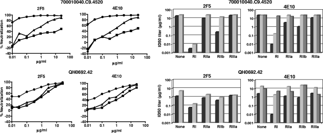
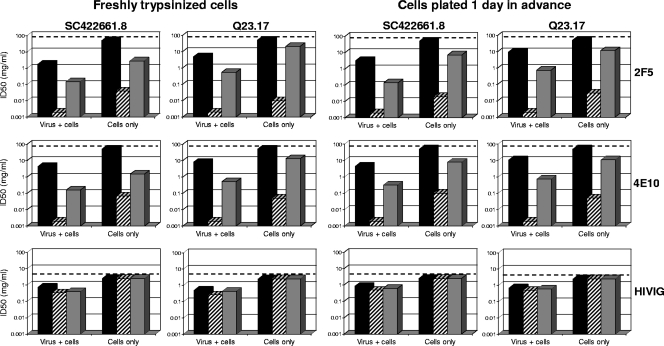
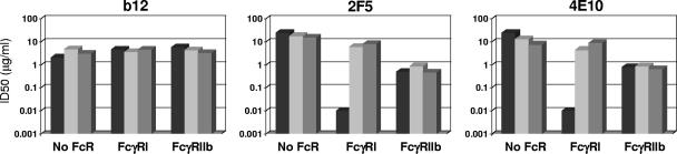
Receptors (FcgammaRs) for the constant region of immunoglobulin G (IgG) are an important link between humoral immunity and cellular immunity. To help define the role of FcgammaRs in determining the fate of human immunodeficiency virus type 1 (HIV-1) immune complexes, cDNAs for the four major human Fcgamma receptors (FcgammaRI, FcgammaRIIa, FcgammaRIIb, and FcgammaRIIIa) were stably expressed by lentiviral transduction in a cell line (TZM-bl) commonly used for standardized assessments of HIV-1 neutralization. Individual cell lines, each expressing a different FcgammaR, bound human IgG, as evidence that the physical properties of the receptors were preserved. In assays with a HIV-1 multisubtype panel, the neutralizing activities of two monoclonal antibodies (2F5 and 4E10) that target the membrane-proximal external region (MPER) of gp41 were potentiated by FcgammaRI and, to a lesser extent, by FcgammaRIIb. Moreover, the neutralizing activity of an HIV-1-positive plasma sample known to contain gp41 MPER-specific antibodies was potentiated by FcgammaRI. The neutralizing activities of monoclonal antibodies b12 and 2G12 and other HIV-1-positive plasma samples were rarely affected by any of the four FcgammaRs. Effects with gp41 MPER-specific antibodies were moderately stronger for IgG1 than for IgG3 and were ineffective for Fab. We conclude that FcgammaRI and FcgammaRIIb facilitate antibody-mediated neutralization of HIV-1 by a mechanism that is dependent on the Fc region, IgG subclass, and epitope specificity of antibody. The FcgammaR effects seen here suggests that the MPER of gp41 could have greater value for vaccines than previously recognized.


Figures

FIG. 1.
FIG. 1.
Expression of the FcγRs on TZM-bl cells. Cell surface expression of the Fcγ receptors was analyzed by flow cytometry using the four established cell lines. Cell suspensions were stained with FITC-labeled mouse monoclonal Abs against the human FcγRs at 4°C. After washing, cells were fixed and analyzed using a BD FACSCalibur analyzer (BD Biosciences). The acquired data were analyzed using a FlowJo software (Tree Star). (A) Cells transduced with FcγRI cDNA stained with anti-human CD64. (B) Cells transduced with FcγRIIa cDNA stained with anti-human CD32. (C) Cells transduced with FcγRIIb stained with anti-human CD32. (D) Cells transduced with FcγRIIIa cDNA stained with anti-human CD16. (E) Cells transduced with FcγRI cDNA stained with anti-human FcɛR-γ chain. (F) Cells transduced with FcγRIIIa cDNA stained with anti-γ-chain. Black-filled curves correspond to nonstained cells; gray-filled curves correspond to cells stained with isotype control Abs.
FIG. 2.
FIG. 2.
Binding of human IgG subclasses to TZM-bl cells expressing different FcγRs. Pure myeloma proteins of the four IgG subclasses were incubated with a suspension of each of the FcγR-expressing cells. After washing to remove unbound myeloma proteins, bound proteins were quantified by flow cytometry. Because the medium- to low-affinity receptors FcγRIIa, FcγRIIb, and FcγRIIIa bind mIgG poorly, the myeloma proteins were heat aggregated before use in these specific cases. Nonaggregated myeloma proteins were used for FcγRI-expressing cells.
FIG. 3.
FIG. 3.
Effect of FcγR expression on HIV-1 neutralization by broadly neutralizing monoclonal Abs and HIVIG. The neutralizing activities of monoclonal Abs b12, 2G12, 2F5, and 4E10 (all IgG1) and HIVIG (purified IgG fraction prepared from pooled HIV-1-positive plasma samples) were assessed against four strains each of HIV-1 subtype B (6535.3, QH0692.42, SC05.8C11.2344, and SC422661.87), subtype C (Du156.12, Du422.1, ZM197M.PB7, and CAP45.2.00.G3), and subtype A (Q23.17, Q259.d2.17, Q461.a2, and Q769.d22) in parental TZM-bl cells (black bars) and in TZM-bl cells expressing FcγR (gray bars). ID50, 50% inhibitory dose.
FIG. 4.
FIG. 4.
Effect of FcγR expression on HIV-1 neutralization by plasma samples from infected individuals. The neutralizing activities of plasma samples from individuals who were chronically infected with either HIV-1 subtype C (BB12, BB24, BB34, and BB87) or subtype B (HIV 011, HIV 012, HIV 013, HIV 014, 1648, 1652, and 1686) were assessed against two subtype B viruses (700010040.C94520 and QH0692.42) and two subtype C viruses (CAP45.2.00.G3 and ZM197M.PB7) in parental TZM-bl cells (black bars) and in TZM-bl cells expressing FcγRI (left-hatched bars), FcγRIIa (gray bars), FcγRIIb (right-hatched bars), and FcγRIIIa (horizontally hatched bars). Fifty percent inhibitory dose (ID50) neutralization titers that were below the level of detection (dilution of <1:20) were assigned a value of 10 and are not visible.
FIG. 5.
FIG. 5.
Amino acid sequence alignment of the MPER region of the HIV-1 Env-pseudotyped viruses used in this study. The minimal epitopes for 2F5 and 4E10 are boxed. Key amino acid changes in the 2F5 epitope that are referred to in the text are shown in bold.
FIG. 6.
FIG. 6.
Importance of IgG subclass and the Fc region of Ab. Left panel, neutralization curves for 2F5 (IgG1) and 4E10 (IgG1) assayed against two HIV-1 subtype B Env-pseudotyped viruses in parental TZM-bl cells (squares) and in TZM-bl cells expressing either FcγRI (circles) or FcγRIIb (triangles). Right panel, neutralization potencies of 2F5 IgG1 and 4E10 IgG1 (black bars), 2F5 IgG3 and 4E10 IgG3 (light gray bars), and 4E10 Fab (dark gray bars) in parental TZM-bl cells and TZM-bl cells expressing either FcγRI, FcγRIIa, FcγRIIb, or FcγRIIIa. ID50, 50% inhibitory dose.
FIG. 7.
FIG. 7.
Enhanced neutralizing activities of gp41 MPER-specific Abs captured by FcγRI and FcγRIIb on the cell surface prior to virus exposure. Neutralizing activities of 2F5, 4E10, and HIVIG were assessed against two strains of HIV-1 (subtype B strain SC422661.87 and subtype A strain Q23.17) in parental TZM-bl cells (black bars) and in TZM-bl cells expressing either FcγRI (hatched bars) or FcγRIIb (gray bars). The assays were performed either with freshly trypsinized cells or with preseeded cells that had been incubated for 1 day before the start of the assay. Two assay formats were used in both cases: (i) standard conditions in which virus was incubated with Ab for 1 h prior to addition of the combined mixture to cells (virus + cells) and (ii) a modified format in which cells were preincubated with Ab for 2 h, followed by one complete change of the medium to remove Ab prior to addition of virus (cells only). The highest Ab concentrations tested are indicated by a dashed line; thus, values below this dashed line signify positive neutralization. ID50, 50% inhibitory dose.
FIG. 8.
FIG. 8.
Relative binding of HIV-1 broadly neutralizing monoclonal Abs to FcγRI on TZM-bl cells. TZM-bl cells expressing FcγRI were incubated with either IgG1b12, 2G12, 2F5, or 4E10 for 1 h at 4°C. Cells were washed three times, stained with FITC-conjugated goat anti-human IgG (Fab specific), and analyzed by flow cytometry. Top, percentage of cells staining positive; bottom, mean fluorescence intensity (MFI) of positive cells. Background staining with an isotype control Ab was subtracted in both cases.
FIG. 9.
FIG. 9.
Normal human serum abrogates the FcγRI effect on 2F5 and 4E10. Neutralization assays were performed with HIV-1 700010040.C9.4520 in parental TZM-bl cells and in TZM-bl cells expressing either FcγRI or FcγRIIb in the absence of serum (black bars) or in the presence of either 5% fresh normal human serum (light gray bars) or 5% heat-inactivated normal human serum (dark gray bars). The monoclonal Abs were of the IgG1 subclass. ID50, 50% inhibitory dose.

References

    1. Alam, S. M., M. McAdams, D. Boren, M. Rak, R. M. Scearce, F. Gao, Z. T. Camacho, D. Gewirth, G. Kelsoe, P. Chen, and B. F. Haynes. 2007. The role of antibody polyspecificity and lipid reactivity in binding of broadly neutralizing anti-HIV-1 envelope human monoclonal antibodies 2F5 and 4E10 to glycoprotein 41 membrane proximal envelope epitopes. J. Immunol. 1784424-4435. - PMC - PubMed
    1. Baravalle, G., M. Brabec, L. Snyers, D. Blaas, and R. Fuchs. 2004. Human rhinovirus type 2-antibody complexes enter and infect cells via Fcγ receptor IIB1. J. Virol. 782729-2737. - PMC - PubMed
    1. Barbato, G., E. Bianchi, P. Ingallinella, W. H. Hurni, M. D. Miller, G. Ciliberto, R. Cortese, R. Bazzo, J. W. Shiver, and A. Pessi. 2003. Structural analysis of the epitope of the anti-HIV antibody 2F5 sheds light into its mechanism of neutralization and HIV fusion. J. Mol. Biol. 3301101-1115. - PubMed
    1. Barnes, N., A. L. Gavin, P. S. Tan, P. Mottram, F. Koentgen, and P. M. Hogarth. 2002. FcγRI-deficient mice show multiple alterations to inflammatory and immune responses. Immunity 16379-389. - PubMed
    1. Baum, L. L., K. J. Cassutt, K. Knigge, R. Khattri, J. Margolick, C. Rinaldo, C. A. Kleeberger, P. Nishanian, D. R. Henrard, and J. Phair. 1996. HIV-1 gp120-specific antibody-dependent cell-mediated cytotoxicity correlates with rate of disease progression. J. Immunol. 1572168-2173. - PubMed

Publication types