. 2009 Jul 24;284(30):19791-9.
doi: 10.1074/jbc.M109.020362. Epub 2009 May 19.

Modulation of the membrane type 1 matrix metalloproteinase cytoplasmic tail enhances tumor cell invasion and proliferation in three-dimensional collagen matrices

Affiliations

Modulation of the membrane type 1 matrix metalloproteinase cytoplasmic tail enhances tumor cell invasion and proliferation in three-dimensional collagen matrices

Natalie M Moss et al. J Biol Chem. .

Abstract

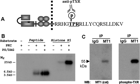
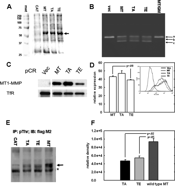
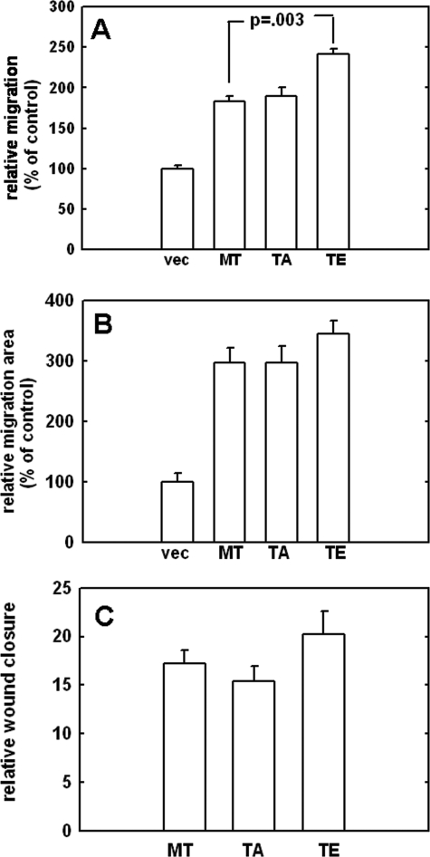
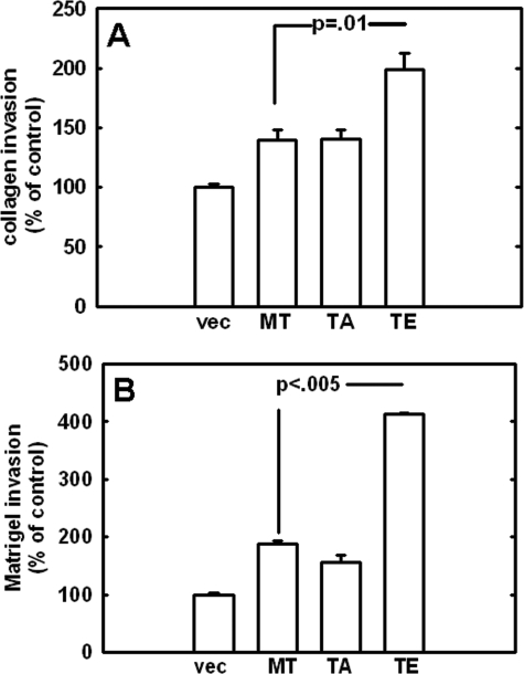
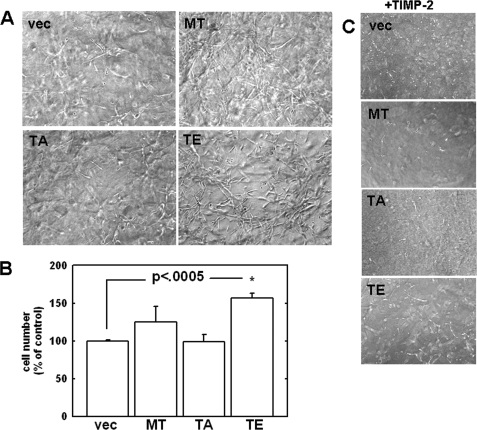
Increasing evidence suggests that the cytoplasmic tail of membrane type 1 matrix metalloproteinase (MT1-MMP) is subject to phosphorylation and that this modification may influence its enzymatic activity at the cell surface. In this study, phosphorylated MT1-MMP is detected using a phospho-specific antibody recognizing a protein kinase C consensus sequence (phospho-TXR), and a MT1-MMP tail peptide is phosphorylated by exogenous protein kinase C. To characterize the potential role of cytoplasmic residue Thr(567) in these processes, mutants that mimic a state of either constitutive (T567E) or defective phosphorylation (T567A) were expressed and analyzed for their functional effects on MT1-MMP activity and cellular behavior. Phospho-mimetic mutants of Thr(567) exhibit enhanced matrix invasion as well as more extensive growth within a three-dimensional type I collagen matrix. Together, these findings suggest that MT1-MMP surface action is regulated by phosphorylation at cytoplasmic tail residue Thr(567) and that this modification plays a critical role in processes that are linked to tumor progression.


Figures

FIGURE 1.
FIGURE 1.
Analysis of MT1-MMP cytoplasmic tail residue Thr567. A, schematic of the MT1-MMP cytoplasmic tail sequence showing the location of Thr567 within the PKC consensus recognition sequence TXR. B, in vitro kinase assays were conducted using a synthetic peptide containing the MT1-MMP tail sequence as substrate and recombinant PKCδ. Reactions were carried out in the presence or absence of purified PKC and were initiated in the presence or absence of diacylglycerol (DAG) and phosphatidylserine (PS), as indicated. Polypeptides were resolved by electrophoresis and analyzed by autoradiography. C, MT1-MMP was immunoprecipitated from whole cell lysates using an antibody recognizing the hinge domain of MT1-MMP. Following electrophoresis and electroblotting, immunoprecipitate (IP) blots were probed with antibody directed against the MT1-MMP catalytic domain (left) or anti-phospho-TXR (right).
FIGURE 2.
FIGURE 2.
Analysis of MT1-MMP Thr567 mutants. A, expression of MT1-MMP constructs. MDA-MB-231 cells were transfected with FLAG epitope-tagged constructs containing either wild type MT1-MMP (MT), empty vector (CAT), or mutations at cytoplasmic residue Thr567 that either mimic (TE) or prevent (TA) phosphorylation. Cells were lysed, and lysates were subjected to Western blotting using anti-FLAG M2 antibodies as described under “Experimental Procedures.” mwt, molecular weight standards in kDa. B, gelatin zymography was performed on serum-free conditioned medium from cells transfected with empty vector (vec), wild type MT1-MMP (MT), or the T567A and T567E mutants (TA and TE, respectively). Controls included cells transfected with wild type MT1-MMP cultured in the presence of the broad spectrum MMP inhibitor GM6001 (MT/GM). Arrows a–c, the migration positions of pro-MMP-2, intermediate MMP-2, and active MMP-2, respectively. C, surface expression of MT1-MMP constructs. Stable cell lines were generated that display comparable levels of MT1-MMP expression in whole cell lysates. To evaluate surface presentation, surface proteins were first biotinylated with a non-cell-permeable biotin analog and lysed, and lysates were precipitated with NeutrAvidin to isolate surface-associated proteins, followed by analysis for MT1-MMP by Western blotting using anti-FLAG M2, as described under “Experimental Procedures.” D, fluorescence-activated cell sorting analysis was conducted using monoclonal M2 FLAG antibody and AlexaTM 488-conjugated anti-mouse IgG secondary antibody to quantify the amount of cell surface MT1-MMP as described under “Experimental Procedures.” E, cells transfected with wild type or mutant MT1-MMP were treated with the serine-threonine phosphatase inhibitor Calyculin A (20 nm, 10 min), and lysates were subjected to immunoprecipitation using anti-phospho-Thr antibody (2 μg). Immunoprecipitates were then subjected to Western blotting with anti-FLAG M2, as described under “Experimental Procedures.” The arrow denotes the migration position of MT1-MMP. *, nonspecific binding. F, densitometric quantitation of triplicate repeat experiments as in E.
FIGURE 3.
FIGURE 3.
Analysis of cell migration. A, the effect of Thr567 mutation on chemotactic migration was assessed using a Boyden chamber migration assay. The underside of the chamber was coated with type I collagen (10 μg/ml). Cells were serum-starved overnight, incubated in Boyden chambers, and allowed to migrate for 3.5 h at 37 °C prior to enumeration of migrating cells. Results are the averages of three independent experiments and are shown as percentages relative to vector controls (designated 100%). B, the colloidal gold migration assay was used to evaluate collagen-driven single cell motility. Cells were serum-starved and allowed to migrate for 12 h prior to quantitation of phagokinetic tracks by computer-assisted image analysis. Results are the averages of three independent experiments and are shown as percentages relative to vector controls (designated 100%). C, the effect of Thr567 mutation on experimental wound closure was assessed using a scratch wound assay. Cells were plated to confluence and serum-starved overnight. Scratches were mechanically introduced to the monolayer using a micropipette tip. Cells were incubated at 37 °C for 18 h. Results are expressed as relative wound closure compared with wound width at time 0.
FIGURE 4.
FIGURE 4.
Analysis of cell invasion. The effect of Thr567 mutation on cell invasion was analyzed using Boyden chambers overlaid with gels composed of type I collagen (A) or Matrigel (B), as described under “Experimental Procedures.” Cells were serum-starved overnight prior to incubation in coated Boyden chambers for 18 h at 37 °C and enumeration of invading cells. Results are the averages of three independent experiments and are shown as percentages relative to vector controls (designated 100%).
FIGURE 5.
FIGURE 5.
Analysis of growth in three-dimensional collagen gels. A, three-dimensional cultures were prepared by diluting type I rat tail collagen with complete medium to a final concentration of 1.5 mg/ml. A, cells (5 × 10⁴) were seeded into the collagen mixture prior to solidification. Gels were allowed to incubate at 37 °C for 8 days and were photographed using phase-contrast microscopy. B, quantitation of proliferation within three-dimensional collagen gels was performed by dissolution of the gels using bacterial collagenase, followed by hemocytometry, as described under “Experimental Procedures” (19). C, in control experiments, the MMP inhibitor TIMP-2 was also included at a final concentration of 5 μg/ml (right), and the experiment was performed as in A.

References

    1. Sternlicht M. D., Werb Z. (2001) Annu. Rev. Cell Dev. Biol. 17, 463–516 - PMC - PubMed
    1. Sabeh F., Ota I., Holmbeck K., Birkedal-Hansen H., Soloway P., Balbin M., Lopez-Otin C., Shapiro S., Inada M., Krane S., Allen E., Chung D., Weiss S. J. (2004) J. Cell Biol. 167, 769–781 - PMC - PubMed
    1. Sato H., Takino T., Okada Y., Cao J., Shinagawa A., Yamamoto E., Seiki M. (1994) Nature 370, 61–65 - PubMed
    1. Buccione R., Orth J. D., McNiven M. A. (2004) Nat. Rev. Mol. Cell Biol. 5, 647–657 - PubMed
    1. Jiang A., Lehti K., Wang X., Weiss S. J., Keski-Oja J., Pei D. (2001) Proc. Natl. Acad. Sci. U.S.A. 98, 13693–13698 - PMC - PubMed

Publication types

MeSH terms