. 2009 Jul;150(3):1219-34.
doi: 10.1104/pp.109.138388. Epub 2009 May 20.

AtBXL1 encodes a bifunctional beta-D-xylosidase/alpha-L-arabinofuranosidase required for pectic arabinan modification in Arabidopsis mucilage secretory cells

Affiliations

AtBXL1 encodes a bifunctional beta-D-xylosidase/alpha-L-arabinofuranosidase required for pectic arabinan modification in Arabidopsis mucilage secretory cells

Andrej A Arsovski et al. Plant Physiol. 2009 Jul.

Abstract

Following pollination, the epidermal cells of the Arabidopsis (Arabidopsis thaliana) ovule undergo a complex differentiation process that includes the synthesis and polar secretion of pectinaceous mucilage followed by the production of a secondary cell wall. Wetting of mature seeds leads to the rapid bursting of these mucilage secretory cells to release a hydrophilic gel that surrounds the seed and is believed to aid in seed hydration and germination. A novel mutant is identified where mucilage release is both patchy and slow and whose seeds display delayed germination. While developmental analysis of mutant seeds reveals no change in mucilage secretory cell morphology, changes in monosaccharide quantities are detected, suggesting the mucilage release defect results from altered mucilage composition. Plasmid rescue and cloning of the mutant locus revealed a T-DNA insertion in AtBXL1, which encodes a putative bifunctional beta-d-xylosidase/alpha-l-arabinofuranosidase that has been implicated as a beta-d-xylosidase acting during vascular development. Chemical and immunological analyses of mucilage extracted from bxl1 mutant seeds and antibody staining of developing seed coats reveal an increase in (1-->5)-linked arabinans, suggesting that BXL1 is acting as an alpha-l-arabinofuranosidase in the seed coat. This implication is supported by the ability to rescue mucilage release through treatment of bxl1 seeds with exogenous alpha-l-arabinofuranosidases. Together, these results suggest that trimming of rhamnogalacturonan I arabinan side chains is required for correct mucilage release and reveal a new role for BXL1 as an alpha-l-arabinofuranosidase acting in seed coat development.


Figures

Figure 1.
Figure 1.
Ruthenium red staining and seed coat structure of bxl1-1 versus wild-type Ws seeds. A and B, Wild-type (A) and bxl1-1 (B) seeds shaken in water and then stained with ruthenium red. Note patchy mucilage staining around bxl1-1 seeds. C and D, Wild-type (C) and bxl1-1 (D) seeds stained with ruthenium red after shaking in 0.05 m EDTA. E to L, Cross sections of developing seed coat epidermal cells stained with toluidine blue. E to H, Wild type. E, 4 DPA with central vacuole filling most of cell. F, 7 DPA, purple-staining mucilage is accumulating. G, 10 DPA, dark purple-staining mucilage found in upper tangential regions of the cell, above the blue-staining secondary cell wall forming around the cytoplasm. H, 13 DPA, secondary cell wall (blue) has filled in the central region of the cell. I to L, bxl1-1 mutant sections, note their similarity at each stage to the wild type. I, 4 DPA; J, 7 DPA; K, 10 DPA; L, 13 DPA. Bars = 200 μm in A to D and 50 μm in E to L.
Figure 2.
Figure 2.
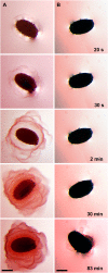
Time lapse of mucilage release for bxl1-1 versus wild-type Ws seeds. Seeds were placed in ruthenium red solution and photographed over 90 min. A, Wild-type seed releases mucilage within 20 s and then mucilage stains pink over time. B, bxl1-1 seed shows no mucilage release until 83 min, at which time mucilage is observed only for a patch of cells. Bars = 100 μm. [See online article for color version of this figure.]
Figure 3.
Figure 3.
Germination of bxl1-1 versus Ws (WS) wild-type seeds. Germination of seeds placed on minimal medium agar plates and cold treated for 72 h. Seeds were either plated directly as dry seeds (no trtmt) or prehydrated by shaking for 90 min in water (H2O) or 0.05 m EDTA as indicated, followed by suspension in 0.01% agarose and plating on minimal medium. Samples were done in triplicate with 50 to 60 seeds per sample. Error bars represent se.
Figure 4.
Figure 4.
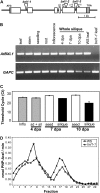
Structure and transcription of AtBXL1. A, Illustration of the intron-exon structure of AtBXL1 showing the location of the different T-DNA insertions in bxl1-1, bxl1-2, and bxl1-3. Exons are drawn as boxes with gray shading indicating untranslated regions. T-DNA insertions are indicated by triangles. Locations of the primers used for the RT-PCR results shown in B and C are indicated with arrows. B, RT-PCR (30 cycles) using AtBXL1 primers p15/16 on RNA isolated from Col wild-type leaves, stems, seedlings, roots, inflorescence tips, intact siliques at 4, 7, and 10 DPA, plus leaf tissue from wild-type Ws (WS) and bxl1-1 plants. The loading control is cytosolic glyceraldehyde-3-P dehydrogenase (GAPC). C, Real-time PCR using AtBXL1 primers p3/4 on inflorescences (inflo), intact 4-DPA siliques (sd + sil), and separated seeds and siliques at 7 and 10 DPA. Duplicate samples were performed using GAPC primers and used to normalize the AtBXL1 results. Error bars represent se, n = 3. D, Analysis of the α-l-arabinofuranosidase activity in bxl1-1 mutant versus wild-type siliques using cation-exchange chromatography. Protein extracts were analyzed by SP-Sepharose chromatography and collected as 1-mL fractions. Each fraction was assayed for α-l-arabinofuranosidase activity and normalized against β-galactosidase activity from the same fraction.
Figure 5.
Figure 5.
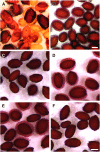
Molecular complementation with PTYg and phenotypic rescue of bxl1-1 mutants with exogenous enzymes. A and B, Molecular complementation results demonstrating restoration of mucilage release in bxl1-1 PTYg transformants (B) and patchy phenotype of bxl1-1 pGREEN 0229-transformed control lines (A). C to F, Treatment of bxl1-1 with exogenous enzymes. C, Enzyme buffer control; note patchy mucilage release. D and E, Treatment with α-l-arabinofuranosidases AN1571 (D) and AN7908 (E); note almost wild-type mucilage levels in (D) and significantly increased mucilage release in (E) compared to (C) and (F). F, Treatment with β-d-xylosidase AN2359; note patchy mucilage release. Bars = 200 μm.
Figure 6.
Figure 6.
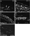
Immunofluorescence of developing Ws (WS) versus bxl1-1 seeds coats. A and B, Confocal sections of 7-DPA Ws and bxl1-1 seed coats stained with anti-arabinan antibody LM6. Arrows indicate lower corners of the mucilage cells; note intense staining around whole cell in bxl1-1 mutants. C and D, Confocal sections of 7-DPA Ws and bxl1-1 seed coats stained with unbranched RG I antibody CCRC-M36. E, Control image of Ws seed coat stained without primary antibody. Photo contrast was enhanced to visualize cells. All photographs were taken with the same microscope settings, save B, where the gain had to be reduced due to saturation of the image due to high intensity of fluorescence, making the image in B an underrepresentation of the degree of labeling. Bar = 50 μm.
Figure 7.
Figure 7.
Phenotype of bxl1-1 double mutants with ap2-1, ttg1-1, myb61-1, and mum4-1. Ruthenium red staining of single mutants and double mutants with bxl1-1, all shaken in 0.05 m EDTA to promote release of their small amount of mucilage prior to staining, except myb61-1 and bxl1-1 myb61-1 (E and F), which were shaken in water since myb61-1 releases mucilage in water. A and B, ap2-1 and bxl1-1 ap2-1, respectively. C and D, ttg1-1 and bxl1-1 ttg1-1, respectively. E and F, myb61-1 and bxl1-1 myb61-1, respectively; note mucilage release in myb61-1 but not in the double mutant. G and H, mum4-1 and bxl1-1 mum4-1, respectively; note thin layer of mucilage surrounding mum4-1 seeds that is not seen in the double mutant. Bars = 500 μm.

References

    1. Alonso J, Stepanova AN, Leisse TJ, Kim CJ, Zimmerman J, Barajas P, Cheuk R, Gadrinab C, Heller C, Jeske A (2003) Genome-wide insertional mutagenesis of Arabidopsis thaliana. Science 301 653–657 - PubMed
    1. Bauer S, Vasu P, Mort AJ, Somerville CR (2005) Cloning, expression, and characterization of an oligoxyloglucan reducing end-specific xyloglucanbiohydrolase from Aspergillus nidulans. Carbohydr Res 340 2590–2597 - PubMed
    1. Bauer S, Vasu P, Persson S, Mort AJ, Somerville CR (2006) Development and application of a suite of polysaccharide-degrading enzymes for analyzing plant cell walls. Proc Natl Acad Sci USA 103 11417–11422 - PMC - PubMed
    1. Beeckman T, De Rycke R, Viane R, Inzé D (2000) Histological study of seed coat development in Arabidopsis thaliana. J Plant Res 113 139–148
    1. Carpita N, Gibeaut DM (1993) Structural models of primary cell walls in flowering plants: consistency of molecular structure with the physical properties of the walls during growth. Plant J 3 1–30 - PubMed

Publication types

MeSH terms