. 2009 Sep;90(Pt 9):2147-56.
doi: 10.1099/vir.0.012047-0. Epub 2009 May 20.

Parainfluenza virus 5 genomes are located in viral cytoplasmic bodies whilst the virus dismantles the interferon-induced antiviral state of cells

Affiliations

Parainfluenza virus 5 genomes are located in viral cytoplasmic bodies whilst the virus dismantles the interferon-induced antiviral state of cells

T S Carlos et al. J Gen Virol. 2009 Sep.

Abstract

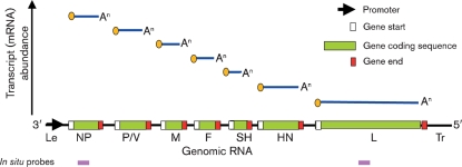
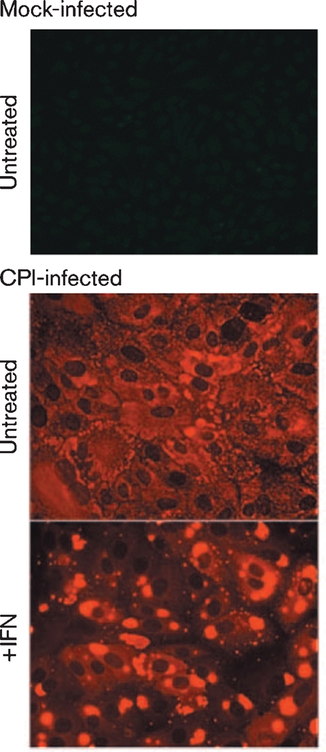
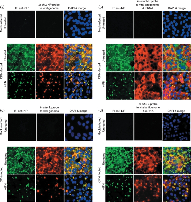
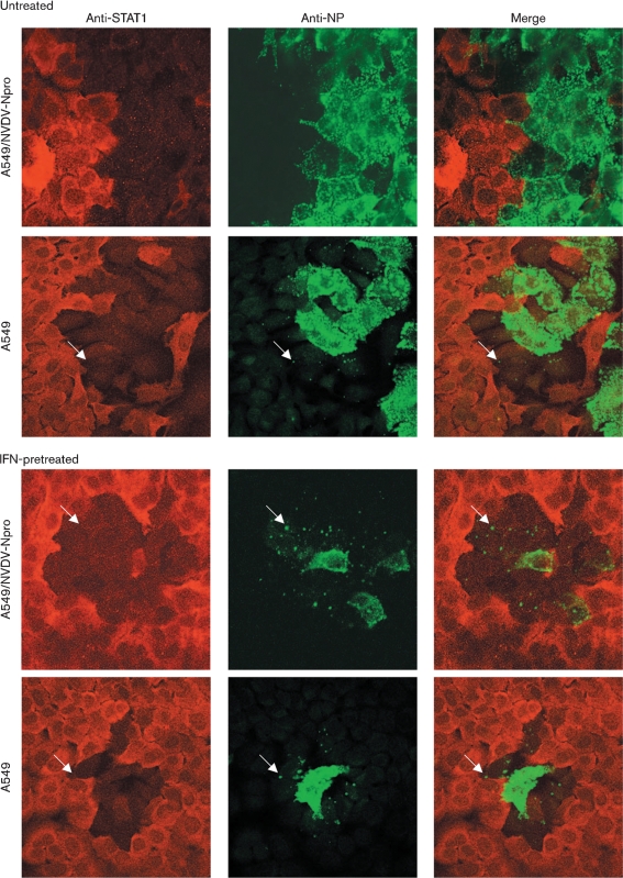
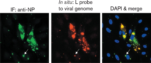
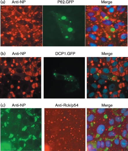
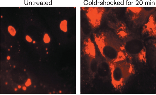
Although the replication cycle of parainfluenza virus type 5 (PIV5) is initially severely impaired in cells in an interferon (IFN)-induced antiviral state, the virus still targets STAT1 for degradation. As a consequence, the cells can no longer respond to IFN and after 24-48 h, they go out of the antiviral state and normal virus replication is established. Following infection of cells in an IFN-induced antiviral state, viral nucleocapsid proteins are initially localized within small cytoplasmic bodies, and appearance of these cytoplasmic bodies correlates with the loss of STAT1 from infected cells. In situ hybridization, using probes specific for the NP and L genes, demonstrated the presence of virus genomes within these cytoplasmic bodies. These viral cytoplasmic bodies do not co-localize with cellular markers for stress granules, cytoplasmic P-bodies or autophagosomes. Furthermore, they are not large insoluble aggregates of viral proteins and/or nucleocapsids, as they can simply and easily be dispersed by 'cold-shocking' live cells, a process that disrupts the cytoskeleton. Given that during in vivo infections, PIV5 will inevitably infect cells in an IFN-induced antiviral state, we suggest that these cytoplasmic bodies are areas in which PIV5 genomes reside whilst the virus dismantles the antiviral state of the cells. Consequently, viral cytoplasmic bodies may play an important part in the strategy that PIV5 uses to circumvent the IFN system.


Figures

Fig. 1.
Fig. 1.
Schematic diagram of the gene order and the transcript abundance of PIV5 mRNAs (see text for details). The positions on the genome map that the NP and L in situ probes bind to are shown by pink boxes.
Fig. 2.
Fig. 2.
Visualization of viral cytoplasmic bodies in CPI-infected cells treated with IFN. Vero cell monolayers were infected with CPI at a high m.o.i. (50–100 p.f.u. per cell). After an adsorption period of 1–2 h on a rocking platform at 37 °C, the virus inoculum was removed and replaced with fresh maintenance medium. At 12 h p.i., the medium was either supplemented with rHuIFN-α or left untreated as a negative control. At 36 h post treatment, the cells were fixed and the distribution of the NP was visualized by immunofluorescence using a Nikon Microphot-FXA immunofluorescence microscope.
Fig. 3.
Fig. 3.
Detection of genomic RNA and antigenomic RNA/mRNA in CPI-infected cells. Vero cells were either mock-infected or infected with CPI at a high m.o.i., and at 8 h p.i., the cells were or were not treated with IFN. At 48 h p.i., the cells were fixed and co-stained by immunofluorescence, with an antibody to NP, and by in situ hybridization, using probes specific for genomic NP (a) or L (c) RNA or NP (b) or L (d) antigenomic RNA/mRNA. The cells were also counter-stained with DAPI to reveal the location of the nuclei. The final column in all four panels is the merged patterns from all three stains. Cells were visualized using a Leica DM5000B wide-field fluorescence microscope. Note: NP and L probes that bind to the genome should give the same intensity of staining, whilst the NP probe that binds to mRNA/antigenomes should give more intense staining than the L probe that binds to mRNA/antigenomes as the abundance of the NP mRNA is significantly greater than that of the L mRNA (see Fig. 1). IF, Immunofluorescence.
Fig. 4.
Fig. 4.
Plaques of PIV5 strain W3A formed on monolayers of MRC5, MRC5/BVDV-Npro, A549 and A549/BVDV-Npro. Note MRC/5BVDV-Npro and A549/BVDV-Npro cells cannot produce IFN in response to virus infection as BVDV-Npro targets IRF-3 for degradation (Hilton et al., 2006). MRC5 cells were fixed at 6 days p.i., whilst the A549 cells were fixed at 10 days p.i.; both were immunostained with an antibody to PIV5 NP.
Fig. 5.
Fig. 5.
A549 and A549/BVDV-Npro cells were or were not pretreated with IFN for 18 h prior to infection with W3A at an m.o.i. of 0.01 p.f.u. per cell. At 4 days p.i., the cells were fixed and co-immunostained for STAT1 and PIV5 NP. Cells were visualized using a Zeiss LSM 5 Exciter confocal microscope. Arrows highlight cells at the edge of the plaque in which small viral cytoplasmic bodies can be detected and in which STAT1 has been degraded. A large, high-resolution copy of this image is available in JGV Online.
Fig. 6.
Fig. 6.
Detection of viral genomes in PIV5 cytoplasmic bodies in cells on the periphery of a developing plaque as PIV5 (W3A) spreads through a monolayer of cells in an IFN-induced antiviral state. Vero cells were infected with W3A at an m.o.i. of 0.01 p.f.u. per cell. At 8 h p.i., IFN was added to the culture medium and, at 48 h p.i., the cells were fixed; the presence of NP was detected by immunofluorescence and genomic RNA was detected using a probe specific for L gene sequences as described in the legend to Fig. 2. Note the presence of low numbers of small viral cytoplasmic bodies (an example of which is indicated with arrows) in the cells surrounding the two cells in which large amounts of viral NP proteins and genomic RNA can be detected. Cells were visualized using a Leica DM5000B wide-field fluorescence microscope. IF, Immunofluorescence.
Fig. 7.
Fig. 7.
Viral cytoplasmic bodies do not co-localize with autophagosomes, cellular cytoplasmic P-bodies or stress granules. Vero cells were transfected with plasmids expressing GFP-tagged marker proteins for autophagosomes (a; P62.GFP) or P-bodies (b; DCP1.GFP). At 24 h post-transfection, the cells were infected with CPI and 12 h later, they were treated with IFN. At 18 h post-treatment, the cells were fixed and immunostained for NP. (c) Untransfected cells were infected and treated with IFN as above and co-stained with an anti-Rck/p54 antibody and an antibody to PIV5 NP. Cells were visualized using a Nikon Microphot-FXA immunofluorescence microscope. No co-localization of the marker proteins and viral proteins was observed.
Fig. 8.
Fig. 8.
Viral cytoplasmic bodies are not large insoluble aggregates of viral proteins. Vero cells were infected with PIV5 CPI at a high m.o.i. and were treated with IFN at 8 h p.i. At 48 h p.i., the cells were or were not cold-shocked for 20 min with ice-cold PBS, after which time the cells were fixed and the distribution of the PIV5 NP visualized by immunofluorescence using a Nikon Microphot-FXA immunofluorescence microscope.

References

    1. Al-Fageeh, M. B. & Smales, C. M. (2006). Control and regulation of the cellular responses to cold shock: the responses in yeast and mammalian systems. Biochem J 397, 247–259. - PMC - PubMed
    1. Andrejeva, J., Childs, K. S., Young, D. F., Carlos, T. S., Stock, N., Goodbourn, S. & Randall, R. E. (2004). The V proteins of paramyxoviruses bind the IFN-inducible RNA helicase, mda-5, and inhibit its activation of the IFN-β promoter. Proc Natl Acad Sci U S A 101, 17264–17269. - PMC - PubMed
    1. Baumgartner, W., Krakowka, S. & Blakeslee, J. R. (1987). Persistent infection of Vero cells by paramyxoviruses. A morphological and immunoelectron microscopic investigation. Intervirology 27, 218–223. - PubMed
    1. Beckham, C. J. & Parker, R. (2008). P bodies, stress granules, and viral life cycles. Cell Host Microbe 3, 206–212. - PMC - PubMed
    1. Bjorkoy, G., Lamark, T., Brech, A., Outzen, H., Perander, M., Overvatn, A., Stenmark, H. & Johansen, T. (2005). p62/SQSTM1 forms protein aggregates degraded by autophagy and has a protective effect on huntingtin-induced cell death. J Cell Biol 171, 603–614. - PMC - PubMed

Publication types

MeSH terms