. 2009 Jul;20(14):3342-52.
doi: 10.1091/mbc.e09-04-0288. Epub 2009 May 20.

Gap junction turnover is achieved by the internalization of small endocytic double-membrane vesicles

Affiliations

Gap junction turnover is achieved by the internalization of small endocytic double-membrane vesicles

Matthias M Falk et al. Mol Biol Cell. 2009 Jul.

Abstract

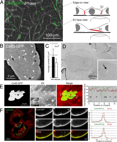
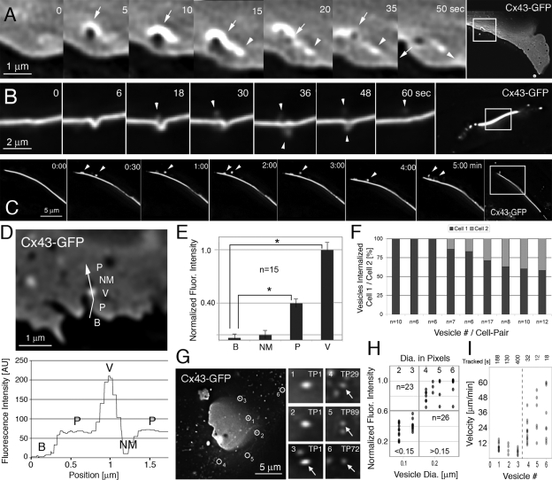
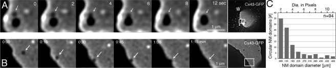
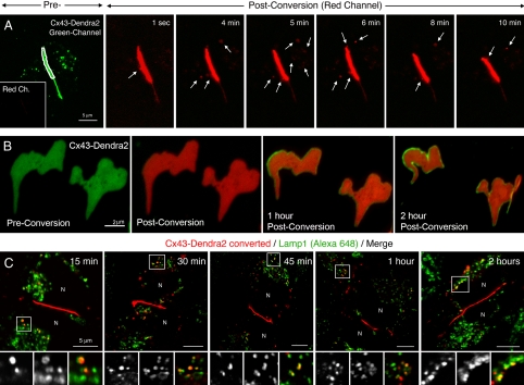
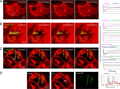
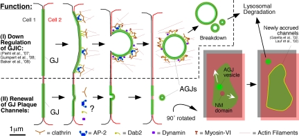
Double-membrane-spanning gap junction (GJ) channels cluster into two-dimensional arrays, termed plaques, to provide direct cell-to-cell communication. GJ plaques often contain circular, channel-free domains ( approximately 0.05-0.5 mum in diameter) identified >30 y ago and termed nonjunctional membrane (NM) domains. We show, by expressing the GJ protein connexin43 (Cx43) tagged with green fluorescent protein, or the novel photoconvertible fluorescent protein Dendra2, that NM domains appear to be remnants generated by the internalization of small GJ channel clusters that bud over time from central plaque areas. Channel clusters internalized within seconds forming endocytic double-membrane GJ vesicles ( approximately 0.18-0.27 mum in diameter) that were degraded by lysosomal pathways. Surprisingly, NM domains were not repopulated by surrounding channels and instead remained mobile, fused with each other, and were expelled at plaque edges. Quantification of internalized, photoconverted Cx43-Dendra2 vesicles indicated a GJ half-life of 2.6 h that falls within the estimated half-life of 1-5 h reported for GJs. Together with previous publications that revealed continuous accrual of newly synthesized channels along plaque edges and simultaneous removal of channels from plaque centers, our data suggest how the known dynamic channel replenishment of functional GJ plaques can be achieved. Our observations may have implications for the process of endocytic vesicle budding in general.


Figures

Figure 1.
Figure 1.
GJ plaques contain circular nonfluorescent domains and juxtaposed bright fluorescent vesicles. (A) HeLa cells expressing Cx43-GFP efficiently assemble GJs in their PMs. GJ plaques can be oriented perpendicular, or horizontally to the image plane (circled). (B) At high light-microscopic resolution, numerous circular, nonfluorescent domains, probably void of GJ channels (dark, arrows), and a comparable number of nearby bright fluorescent vesicles (arrowheads) are detectable in images of horizontally oriented GJ plaques. (C) Quantitative analysis of 7 GJ plaques viewed en face. Large standard deviations indicate that numbers of nonfluorescent domains and juxtaposed vesicles significantly varies between plaques (see Table 1). (D) Circular membrane domains, void of GJ channels, also are detectable by ultrastructural analyses and were termed NM domains or particle-free zones (arrow), as shown, e.g., in a freeze-fracture replica of a GJ plaque derived from fetal rat epidermis. (E and F) GJ plaques and nonjunctional membrane domains contain PM, as indicated by staining with the lipophilic red dye DiI. Cx43-GFP–expressing HeLa cells were stained with DiI (5 μM; 10 min) 16 h after transfection, mounted in a live-cell chamber and imaged immediately after by confocal microscopy. (E) Fluorescence intensities (in arbitrary units) of Cx43-GFP fluorescence (green) and DiI (red) measured along a line traversing PM outside GJs (M), GJ plaques (P), and NM domains of a horizontally oriented GJ plaque. Full-frame image and selected cropped area are shown in the insert. (F) Three sections (z1–z3) spaced 0.2 μm apart through a NM domain (arrow) of a perpendicular-oriented GJ plaque. Quantitative fluorescence intensities of DiI (red) and GFP (green) measured along lines placed through the nonjunctional membrane domain (1), and the GJ plaque (2) are shown on the right. Note the less intense Cx43-GFP fluorescence in the area of the nonjunctional membrane domain in section z2 (top), whereas DiI staining in this area appears at least as intense as in the GJ plaque.
Figure 2.
Figure 2.
Circular nonfluorescent domains appear to be generated by the internalization of small GJ vesicles that bud continuously from GJ plaques. (A–C) High-resolution time-lapse recordings of Cx43-GFP GJ plaques acquired at relatively fast acquisition rates (5 s in A and 6 s in B) reveal the internalization of small vesicles (arrowheads) that bud within seconds from central plaque areas. Vesicle budding appears to generate the circular, nonfluorescent membrane domains (dark, arrows) described above that reside for some time within the plaques and that appear to be preceded by an uprising of the plaque indicated by a strong increase of fluorescence around the bud site. NM domain and vesicle move away from their site of generation (indicated by the position of arrow and arrowhead in the 50-s frame). Vesicles in B appear to bud into both coupled cells, and only into one cell in C. Note the similarity between vesicles shown in Figures 1B and 2C and how vesicles in 2C appear to move along the plaque surface before translocating into the cytoplasm. Vesicles in 2B appear less fluorescent and blurred due to out-of-focus location. Representative magnified frames from Supplemental Movies 2–4 and corresponding full-frame images are shown. (D) Fluorescence intensity (in arbitrary units) measured along a line traversing background area outside a GJ plaque (B), GJ plaque (P), a presumptive internalized GJ vesicle located in front of the plaque (V), and a juxtaposed nonfluorescent membrane domain (NM). The corresponding intensity profile is shown below. (E) Cumulative analyses of 15 comparable line-scans with vesicles located in front of plaques (8) or in front of background (7) (*p < 0.001). Fluorescence of dispersed connexons potentially present in the PM around plaques was not detected. (F) Quantitative analysis of vesicles (n = 77) budding from nine edge-on viewed GJ plaques (≥6 vesicles/plaque) indicates that vesicles preferentially bud into one of two coupled cells; however, with a highly variable bias (58–100%). (G–I) Fluorescence intensity (G and H) and velocity (G and I) measurements indicate that two significantly different populations of Cx43-GFP–containing vesicles are present in the vicinity of GJ plaques: 1) small, bright fluorescent vesicles (probably endocytic; see Results) that moved relatively slowly and interrupted by stationary phases (grouped as >0.15 in H, vesicles 1–3 in G and I, and additional vesicles on the right in H); and 2) even smaller, less fluorescent vesicles (probably secretory; see Results) that moved relatively fast and continuously (grouped as <0.15 in H; vesicles 4–6 in G and I, and additional vesicles on the left in H). The positions of the six vesicles tracked in G and I are circled in the first image (TP1) of the time-lapse sequence and in Supplemental Movie 5. Time points when vesicles first occur are indicated. Dots indicate vesicles in H and distances traveled between two successive frames in I. Darker dots correspond to the overlapping of multiple observations at each position. p values comparing vesicle groups in H and I were <0.001.
Figure 3.
Figure 3.
(A) Optical sections spaced 100 nm apart through a forming vesicle (∼0.7 μm in diameter) that buds from a Cx43-GFP GJ plaque in a fixed transfected HeLa cell. Note the “neck” (arrow) that connects the vesicle with the GJ plaque. (B–H) Ultrastructural analyses of thin-sectioned HeLa cells transfected with Cx43-GFP. (B) Vesicles of various size and shape are associated with a GJ plaque that is recognizable by its typical penta-laminar staining pattern. (C–H) Double-membrane GJ vesicles (with visible penta-laminar staining pattern in F–H) ∼100–200 nm in diameter (arrowheads) at various stages of internalization (C and D, plaque invagination; E and F, attached vesicles; and G and H, released vesicles). Note the electron dense material visible at the neck of attached vesicles (arrows in E and F).
Figure 4.
Figure 4.
Circular nonfluorescent domains can fuse and are expelled at GJ plaque edges. (A and B) Representative still images from Supplemental Movie 6 showing domain fusion (A) and Supplemental Movie 8 showing domain expelling (B). Magnified frames and full-frame images are shown. Nonfluorescent domains (dark) are labeled with arrows. (C) Quantitative size analysis of 94 nonjunctional membrane domains of seven plaques.
Figure 5.
Figure 5.
Photoconversion and tracking of Cx43-Dendra2 GJ plaques allows quantification of vesicle internalization, plaque channel turnover, and degradation by lysosomal pathways. (A) Selected areas restricted to GJ plaques assembled from Cx43-Dendra2 (outlined in white on the green channel preconversion image) were permanently photoconverted to red fluorescence and plaques were followed in the red channel over time (postconversion images). At least 10 vesicles (arrows) budded from the photoconverted plaque area within the 10-min time period shown and moved away from the bud sites into the cytoplasm. Preconversion laser power was set low to prevent accidental photoconversion. No fluorescence signal was detectable in the red channel before photoconversion (inset in A; also see Supplemental Figure S2). (B) Entire GJ plaques were photoconverted, and both channels (green and red) were recorded over time. Within 1 h after conversion, a homogenous green line of GJ channels occurred along the outer plaque edges that widened over time (2 h after conversion), suggesting that newly synthesized, not photoconverted GJ channels accrued along the outer edge of GJs occurring simultaneously with GJ channel internalization. (C) After photoconversion of selected GJ plaques (red), cells were incubated for indicated periods at 37°C, fixed and stained with antibodies directed against the lysosomal marker protein Lamp1, followed by Alexa648-conjugated secondary antibodies (pseudocolored green) and confocal observation. Numerous photoconverted GJ vesicles (red) colocalized at all time points with Alexa648-labeled Lamp1-positive lysosomal structures as indicated by the yellow overlay color. N, nuclei.
Figure 6.
Figure 6.
PM lipids can diffuse throughout GJ plaques, facilitating the generation of NM domains. Cx43-GFP–expressing HeLa cells were labeled for 10 min with the lipophilic dye DiI, as described under Materials and Methods. (A–C) Fluorescence of DiI and Cx43-GFP was photobleached in defined areas in horizontally and perpendicular oriented PM regions, and in GJ plaques (squares in A and B and a circle in C), and fluorescence intensity in photobleached (regions 1 in A and B and regions 1 and 3 in C) and control regions (region 2 in A and B and regions 2 and 4 in C) was measured on time-lapse images acquired at 0.5-s intervals (plotted on the right). DiI staining in the PM and within GJ plaques recovered within a few seconds correlating with a rapid lateral diffusion of PM lipids within and outside GJ plaques. DiI within GJ plaques recovered slower, as indicated by the shallower slope of the DiI recovery curve (red curve in the recovery graph in D), correlating with a less dynamic mobility of PM lipids within GJ plaques. (D) Consistent with a large portion of the membrane area being occupied by protein channels within GJ plaques, amount of DiI within GJ plaques appeared lower than in PM outside GJ plaques (arrow), as indicated by the height of the DiI fluorescence intensity peaks measured along a line-scan performed before photobleaching, and the higher starting point of the fluorescence recovery curve of DiI-photobleached outside GJs (blue curve in the bleach diagram in C, region 3) compared with the recovery curve of DiI photobleached within the GJ (red curve in the bleach diagram in C, region 1). Cx43-GFP did not show detectable recovery within the 8-s time period, and fluorescence intensity of DiI and Cx43-GFP in nonphotobleached control areas remained stable.
Figure 7.
Figure 7.
Schematic representation of the pathways that lead to GJ plaque internalization (described in Piehl et al., 2007; Baker et al., 2008; Gumpert et al., 2008) (I) and GJ plaque channel renewal (described here), GJ vesicle formation (AGJ), and GJ vesicle degradation (II). Whether clathrin and clathrin accessory proteins are involved in the internalization of small GJ vesicles described here has not been determined; however, is probably based on our electron microscopy analyses (see Figure 3). Displacement of nonjunctional membrane domains from central plaque areas (NM domain, gray/red) by lateral movement allows the simultaneous accrual of new channels (yellow) along the periphery of GJ plaques (green) without increasing plaque size consistent with previously published observations (Gaietta et al., 2002; Lauf et al., 2002). Clathrin and accessory proteins are shown in patches in accordance to the appearance of clathrin on GJ plaques (Piehl et al., 2007), and the current thinking that clathrin may provide a scaffold for directed actin assembly, facilitating internalization of large structures such as GJs, viruses, and pathogenic bacteria (Pauly and Drubin, 2007; Veiga et al., 2007). The manner in which clathrin and accessory proteins are drawn remains speculative.

References

    1. Baker S. M., Kim N., Gumpert A. M., Segretain D., Falk M. M. Acute internalization of gap junctions in vascular endothelial cells in response to inflammatory mediator-induced G-protein coupled receptor activation. FEBS Lett. 2008;582:4039–4046. - PMC - PubMed
    1. Beardslee M. A., Laing J. G., Beyer E. C., Saffitz J. E. Rapid turnover of connexin43 in the adult rat heart. Circ. Res. 1998;83:629–635. - PubMed
    1. Berthoud V. M., Minogue P. J., Laing J. G., Beyer E. C. Pathways for degradation of connexins and gap junctions. Cardiovasc. Res. 2004;62:256–267. - PubMed
    1. Bruzzone R., White T. W., Paul D. L. Connections with connexins: the molecular basis of direct intercellular signaling. Eur. J. Biochem. 1996;238:1–27. - PubMed
    1. Bukauskas F. F., Jordan K., Bukauskiene A., Bennett M. V., Lampe P. D., Laird D. W., Verselis V. K. Clustering of connexin 43-enhanced green fluorescent protein gap junction channels and functional coupling in living cells. Proc. Natl. Acad. Sci. USA. 2000;97:2556–2561. - PMC - PubMed

Publication types

Substances