. 2009 Jul;20(14):3239-50.
doi: 10.1091/mbc.e08-12-1255. Epub 2009 May 20.

Regulation of cell polarity through phosphorylation of Bni4 by Pho85 G1 cyclin-dependent kinases in Saccharomyces cerevisiae

Affiliations

Regulation of cell polarity through phosphorylation of Bni4 by Pho85 G1 cyclin-dependent kinases in Saccharomyces cerevisiae

Jian Zou et al. Mol Biol Cell. 2009 Jul.

Abstract

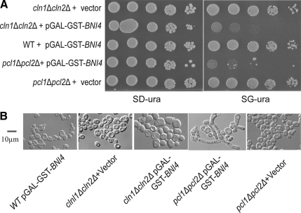
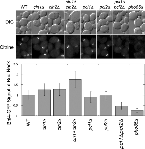
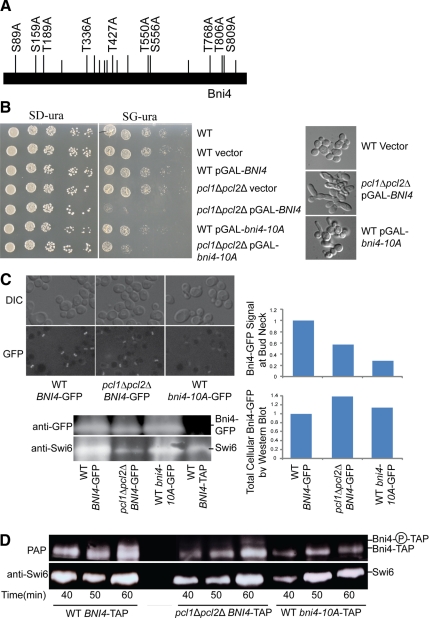
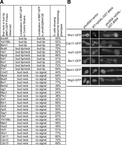
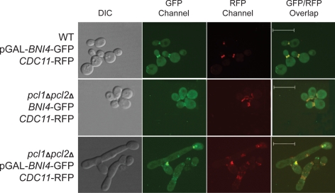
In the budding yeast Saccharomyces cerevisiae, the G1-specific cyclin-dependent kinases (Cdks) Cln1,2-Cdc28 and Pcl1,2-Pho85 are essential for ensuring that DNA replication and cell division are properly linked to cell polarity and bud morphogenesis. However, the redundancy of Cdks and cyclins means that identification of relevant Cdk substrates remains a significant challenge. We used array-based genetic screens (synthetic genetic array or SGA analysis) to dissect redundant pathways associated with G1 cyclins and identified Bni4 as a substrate of the Pcl1- and Pcl2-Pho85 kinases. BNI4 encodes an adaptor protein that targets several proteins to the bud neck. Deletion of BNI4 results in severe growth defects in the absence of the Cdc28 cyclins Cln1 and Cln2, and overexpression of BNI4 is toxic in yeast cells lacking the Pho85 cyclins Pcl1 and Pcl2. Phosphorylation of Bni4 by Pcl-Pho85 is necessary for its localization to the bud neck, and the bud neck structure can be disrupted by overexpressing BNI4 in pcl1Deltapcl2Delta mutant cells. Our data suggest that misregulated Bni4 may bind in an uncontrolled manner to an essential component that resides at the bud neck, causing catastrophic morphogenesis defects.


Figures

Figure 1.
Figure 1.
Genetic interactions with G1 cyclin genes. (A) List of genes that have genetic interactions with G1 cyclin pairs identified from crosses with the deletion strain collection and with a collection of temperature-sensitive strains for synthetic lethal (SL/SS) interactions and with the pGAL-GST-open reading frame (ORF) overexpression collection for synthetic dosage lethal (SDL) interactions. Query genes are listed by row and genetic interactions by column. Synthetic sick interactions are shown in yellow. (B) Pie chart showing the genes that have genetic interactions (either SL/SS or SDL) with G1 cyclin pairs clustered by GO cellular role. Categories that contribute <8% of all genes are grouped as others. (C) Diagram showing one possible scenario for genetic interactions with G1 cyclin pairs. Genes that have SL/SS interaction with CLN1, CLN2 could be in a PCL1, PCL2 regulatory pathway, because quadruple deletion of CLN1, CLN2, PCL1, and PCL2 is lethal, as shown on the left. Genes downstream of PCL1, PCL2 will escape regulation when PCL1 and PCl2 are deleted; therefore, their overexpression may cause toxicity.
Figure 2.
Figure 2.
Synthetic dosage phenotype caused by overexpression of BNI4 in the absence of G1 Cdks. (A) Cultures of WT cells carrying pGAL-GST-BNI4, cln1Δcln2Δ carrying vector (pEGH), cln1Δcln2Δ carrying pGAL-GST-BNI4, pcl1Δpcl2Δ carrying vector (pEGH), and pcl1Δpcl2Δ carrying pGAL-GST-BNI4 were serially diluted and spotted on both SD-ura and SG-ura plates. Plates were incubated 2 d for both SD-ura and SGal-ura plates at 30°C before being photographed. (B) Cultures were grown to log phase in SD-ura, and then transferred to SR-ura and incubated for another 5 h. Galactose was then added to 2%, and cultures were incubated at 30°C for an additional 4.5 h and examined by microscope. Bar, 10 μm.
Figure 3.
Figure 3.
Phosphorylation of Bni4 by Pcl-Pho85 Cdks in vivo and in vitro. (A) Cell cycle Western blot of cultures of BNI4-TAP GAL-CDC20 and pcl1Δpcl2Δ BNI4-TAP GAL-CDC20. Samples were taken from cells at 10-min intervals after release from metaphase arrest for Western blot, FACS analysis, and budding index count. The panel on the top shows Bni4-TAP as detected by PAP. The panel in the middle is the bottom part of the same polyacrylamide gel used for Bni4-TAP detection, but it was probed with anti-Swi6 antibody for a loading control. The bottom panel shows FACS analysis results and budding index counts (% budded cells) for each time point. (B) In vitro phosphorylation of Bni4. C-terminal-GST-tagged Bni4, purified using an E. coli expression system, was mixed with kinase complex consisting of Pcl1 and Pho85; Pcl2 and Pho85; or Cln2, Cdc28, and Cks1; and [γ-32P]ATP in kinase reaction buffer, and incubated at 30°C for 30 min. Reaction products were analyzed by SDS-polyacrylamide gel electrophoresis and autoradiography (left). (C) Pulldown of GST-Bni4 and associated proteins from yeast extracts. Extracts were prepared from strain BY2131 PCL1-myc carrying pGAL-GST-BNI4 in which BNI4 expression had been induced for 3.5 h. Glutathione-Sepharose beads were used to pull down GST-Bni4, and associated proteins were analyzed by Western blot with anti-GST antibody to detect GST-Bni4 and with anti-myc antibody to detect Pcl1-myc. Western blot to detect GST in pull-down samples is not shown.
Figure 4.
Figure 4.
Bni4 localization to the bud neck in Cdk mutant strains. DIC and fluorescence microscopy analysis of Bni4-mCitrine levels at the bud neck in strains of WT, cln1Δ, cln2Δ, cln1Δcln2Δ, pcl1Δ, pcl2Δ, pcl1Δpcl2Δ, and pho85Δ. Cultures were grown to mid-log phase and imaged by fluorescence microscopy. Fluorescence levels at the bud neck were quantified and normalized to wild-type levels. n ≥ 55. Error bars represent SD. Bar, 3 μm.
Figure 5.
Figure 5.
Phenotype associated with Bni4 phosphorylation site mutants. (A) Diagram of Bni4 protein showing all S/TP sites (vertical lines above bar); S/TP sites conserved among members of the Saccharomyces sensu stricto group are shown as raised lines. The S/TP sites substituted with S/TA in bni4-10A are labeled with their amino acid position. (B) Cultures of wild-type strain, wild-type strain containing vector, or GAL-driven plasmid with BNI4, pcl1Δpcl2Δ strain containing vector or a GAL-driven plasmid with BNI4, a wild-type strain containing a GAL-driven plasmid with bni4-10A, and a pcl1Δpcl2Δ strain containing a GAL-driven plasmid with bni4-10A were serially diluted and plated on SD-ura (left) and SG-ura (right, inducing the overexpression of BNI4) and incubated for 2 d at 30°C. To the right are DIC images showing morphology changes in cells in which BNI4 or bni4-10A overexpression has been induced for 4 h. (C) Strains expressing WT BNI4-GFP, pcl1Δpcl2Δ BNI4-GFP, or WT bni4-10A-GFP, with BNI4 under the control of its own promoter, replacing the endogenous gene, were grown in SD medium and Bni4 localization to the bud neck was examined using DIC and fluorescence microscopy. Cultures were grown to mid-log phase. Fluorescence levels at the bud neck were imaged by fluorescence microscopy and quantified by Velocity software, n ≥ 55. Western blot analysis of these cultures is shown at the bottom, along with extract from a BNI4-TAP strain, as a negative control. The top panel was probed with anti-GFP antibody. The bottom panel shows a portion of the same polyacrylamide gel used for Bni4-GFP detection, but was probed with anti-Swi6 antibody as a loading control. The top graph shows relative Bni4-GFP at the bud neck quantified from micrographs as described in Figure 4. The bottom graph shows relative total cellular Bni4-GFP quantified by western blot and normalized to Swi6 levels. (D) Cell-cycle western blot of cultures of pGAL-CDC20 WT BNI4-TAP, pGAL-CDC20 pcl1Δpcl2Δ BNI4-TAP, or pGAL-CDC20 WT bni4-10A-TAP. Samples were analyzed from cultures taken at 10-min intervals from 40 min to 60 min after release from metaphase arrest. The top panel shows Bni4-TAP as detected by PAP complex (PAP). The label Bni4-P-TAP, to the right of the blot, refers to Bni4 phosphoforms, which are undetectable in the bni4-10A-TAP strain. The bottom panel shows a portion of the same polyacrylamide gel used for Bni4-TAP and bni4-10A-TAP detection but was probed with anti-Swi6 antibody as a loading control.
Figure 6.
Figure 6.
Bud neck protein localization in strains overexpressing Bni4. (A) Localization of BNT-(bud neck or bud tip)-GFP fusion proteins observed in control strains, wild-type strains with overexpression of BNI4 or pho85Δ strains carrying vector (second column), or in pho85Δ strains overexpressing BNI4 (third column). Column 4 indicates the percentage of pho85Δ BNT-GFP pGAL-GST-BNI4 cells showing abnormal morphology (i.e., grossly elongated buds and polarity defects). (B) Representative images of cells showing abnormal morphology and mislocalization of BNT-GFP proteins. Overexpression of BNI4 was induced by adding galactose to a log phase culture and incubating at 30°C for 4 h. The GFP signal in these cultures was examined by high-throughput microscopy.
Figure 7.
Figure 7.
Colocalization of Bni4 and septin proteins. WT CDC11-RFP and pcl1Δpcl2Δ CDC11-RFP strains carrying pGAL-BNI4-GFP or vector were grown in medium containing galactose for 4 h and observed using confocal microscopy. Cells were photographed using the DIC, GFP channel, RFP channel, and GFP/RFP overlaid. The RFP signal was switched to red using Volocity software to assist in viewing. Bars, 10 μm.

Comment in

  • Mol Biol Cell. 20:3169.

Similar articles

Cited by

References

    1. Barral Y., Mermall V., Mooseker M. S., Snyder M. Compartmentalization of the cell cortex by septins is required for maintenance of cell polarity in yeast. Mol. Cell. 2000;5:841–851. - PubMed
    1. Barral Y., Parra M., Bidlingmaier S., Snyder M. Nim1-related kinases coordinate cell cycle progression with the organization of the peripheral cytoskeleton in yeast. Genes Dev. 1999;13:176–187. - PMC - PubMed
    1. Boone C., Bussey H., Andrews B. J. Exploring genetic interactions and networks with yeast. Nat. Rev. Genet. 2007;8:437–449. - PubMed
    1. Brachmann C. B., Davies A., Cost G. J., Caputo E., Li J., Hieter P., Boeke J. D. Designer deletion strains derived from Saccharomyces cerevisiae S288C: a useful set of strains and plasmids for PCR-mediated gene disruption and other applications. Yeast. 1998;14:115–132. - PubMed
    1. Cid V. J., Adamikova L., Sanchez M., Molina M., Nombela C. Cell cycle control of septin ring dynamics in the budding yeast. Microbiology. 2001;147:1437–1450. - PubMed

Publication types

MeSH terms