. 2009 Jul;20(14):3390-400.
doi: 10.1091/mbc.e08-12-1172. Epub 2009 May 20.

Scribble interacts with beta-catenin to localize synaptic vesicles to synapses

Affiliations

Scribble interacts with beta-catenin to localize synaptic vesicles to synapses

Yu Sun et al. Mol Biol Cell. 2009 Jul.

Abstract

An understanding of how synaptic vesicles are recruited to and maintained at presynaptic compartments is required to discern the molecular mechanisms underlying presynaptic assembly and plasticity. We have previously demonstrated that cadherin-beta-catenin complexes cluster synaptic vesicles at presynaptic sites. Here we show that scribble interacts with the cadherin-beta-catenin complex to coordinate vesicle localization. Scribble and beta-catenin are colocalized at synapses and can be coimmunoprecipitated from neuronal lysates, indicating an interaction between scribble and beta-catenin at the synapse. Using an RNA interference approach, we demonstrate that scribble is important for the clustering of synaptic vesicles at synapses. Indeed, in scribble knockdown cells, there is a diffuse distribution of synaptic vesicles along the axon, and a deficit in vesicle recycling. Despite this, synapse number and the distribution of the presynaptic active zone protein, bassoon, remain unchanged. These effects largely phenocopy those observed after ablation of beta-catenin. In addition, we show that loss of beta-catenin disrupts scribble localization in primary neurons but that the localization of beta-catenin is not dependent on scribble. Our data supports a model by which scribble functions downstream of beta-catenin to cluster synaptic vesicles at developing synapses.


Figures

Figure 1.
Figure 1.
Scribble localizes to synapses. (A–M) Confocal images of 12 DIV hippocampal cultures. (H–M) To determine the subcellular localization of fluorescently tagged proteins in individual neurons, cells were transfected at <1% efficiency with Lipofectamine 2000 at 10 DIV. Endogenous scribble is localized in a punctate pattern and highly colocalizes with VGLUT-1 (A) and PSD-95 (B) puncta, indicating its localization at excitatory synapses (A–D). Scribble-positive puncta that do not colocalize with VGLUT-1/PSD-95 puncta are also observed (for examples see arrows). Endogenous scribble also colocalizes with synaptophysin (E–G), and Syn-GFP (H–J). RFP-hScrib is localized in a puncta pattern and colocalizes with Syn-GFP (K–M). Scribble-positive puncta that do not colocalize with synaptophysin (arrows), and synaptophysin puncta that do not colocalize with scribble (arrowheads) are observed (E–G). Similarly, the majority of Syn-GFP puncta colocalize with scribble (H–J). Scribble-positive puncta that do not colocalize with Syn-GFP represent immunostaining on cells not transfected with Syn-GFP (H–J; arrows). Arrows indicate RFP-hScrib puncta that do not colocalize with Syn-GFP (K–M). Scale bars, 5 μm.
Figure 2.
Figure 2.
Scribble associates with β-catenin at synapses. (A–E) Confocal images of 12 DIV hippocampal cultures demonstrating colocalization of scribble, β-catenin, and the presynaptic marker, bassoon. Higher magnifications of the inset from A are shown in B–E. The majority of scribble puncta colocalized with β-catenin and bassoon. β-catenin puncta that do not colocalize with scribble (arrows), scribble puncta that do not colocalize with β-catenin (open arrows), and bassoon puncta that do not associate with β-catenin and scribble (arrowheads) are observed. Colocalized scribble and β-catenin puncta that are not associated with bassoon positive sites are also observed (open arrowheads). (F and G) Scribble and β-catenin can be coimmunoprecipitated from synaptosomal fractions. Synaptosomal fractions from E18 brains were immunoprecipitated using β-catenin or scribble antibodies and separated by SDS-PAGE, and immunoblots probed with antibodies specific to scribble, β-catenin, synaptophysin, or cadherin. The input lane corresponds to 40 μg of the crude synaptosomal fraction, P2. Rabbit IgG was used as a control. Scribble and β-catenin were coimmunoprecipitated with one another and with cadherin, but not with the synaptic protein, synaptophysin, indicating the specificity of the immunoprecipitation. N = 2 different preparations. (G) Synaptosomal fractions from E18 brains were affinity purified with GST-β-cat FL, GST-β-cat ΔPDZb, or GST alone bound to glutathione-Sepharose beads. Coomassie blue staining was used to verify the levels of GST and GST fusion proteins used for GST pulldown assay (arrows). Bound proteins were eluted, and blots were probed with anti-scribble or anti-cadherin. Scale bars, 5 μm.
Figure 3.
Figure 3.
RNAi-mediated knockdown of scribble protein levels in primary neurons. (A and B) Neurons were transfected with control RNAi (RNAi-C), or three distinct scribble RNAis (RNAi-1-3) with ∼35% transfection efficiency using the Amaxa nucleofector system. Immunoblot analysis revealed a decrease in scribble protein levels in all three RNAi-expressing cultures, whereas β-catenin levels remained constant. (B) Quantification of immunoblots represents raw data and has not been normalized for transfection efficiency (35.9 ± 2%). N = 3 immunoblots from three separate cultures. *p < 0.05 using Student's t test. (C–L) Confocal images of 10 DIV hippocampal neurons cotransfected using Lipofectamine 2000 (<1% transfection efficiency) with Syn-GFP plus either RNAi-C (D and G) or RNAi-1 (F and J) and immunostained for scribble (C and E) or synaptotagmin (H and K). The neuron expressing RNAi-1 shows a clear reduction in scribble immunostaining in the cell body (E) compared with RNAi-C–expressing cell (C). *, transfected neurons; arrows, untransfected neurons. Syn-GFP is punctate in RNAi-C–expressing cells (D′ and G), but more diffusely localized in cells expressing RNAi-1 (F′ and J). (G–L) Endogenous synaptotagmin is diffusely expressed in RNAi-1–expressing cells. In RNAi-expressing cells Syn-GFP and synaptotagmin are colocalized and display a punctate distribution (G–I). In contrast, in neurons expressing RNAi-1, both Syn-GFP and synaptotagmin are more diffusely distributed (J–L). Synaptotagmin-positive puncta that do not colocalize with Syn-GFP represent immunostaining on cells not transfected with Syn-GFP and RNAi-1. Scale bars, 20 μm.
Figure 4.
Figure 4.
SVs are more diffusely distributed along the axon in neurons expressing scribble RNAi constructs. (A–J) Confocal images of 10 DIV hippocampal neurons cotransfected with Syn-GFP plus scribble RNAis using Lipofectamine 2000 (<1% transfection efficiency) and immunostained for bassoon and PSD-95. Synapses were defined as regions where bassoon and PSD-95 puncta colocalized. (A and F) Images of neurons transfected with RNAi-C (A) and RNAi-1 (F) and pseudocolored for fluorescence intensity. Insets from A and F are shown in higher magnification (B–E and G–J) and selected for histogram analyses (K and L). In RNAi-C–expressing neurons, Syn-GFP is punctate and colocalizes with PSD-95 and bassoon (B–E, arrows). In RNAi-1–expressing neurons, Syn-GFP fluorescence is more diffusely distributed along the axon; however, discrete bassoon and PSD-95 colocalization is still observed (G–J, arrows). Immunopositive bassoon and PSD-95 puncta that do not colocalize with Syn-GFP may represent immunostaining on untransfected neurons. (K and L) Histograms of Syn-GFP fluorescence intensity along a selected axon length in neurons expressing RNAi-C (A and B) or RNAi-1 (F and G) demonstrate that the distribution of Syn-GFP fluorescence is more uniform in scribble knockdown cells and lack distinct punctal and interpunctal regions. (M) Using the Amaxa nucleofector system, neurons were transfected with control RNAi (RNAi-C) or scribble RNAis (RNAi-1-3), and the expression level of synaptophysin and β-catenin was analyzed using Western blot. Despite the fact that synaptophysin protein levels are similar in cultures transfected with RNAi-C and RNAi-1-3 (M), the coverage of Syn-GFP along the axon (the sum of the length of Syn-GFP fluorescence signal per 10 μm axon length ± SE) is greater in RNAi-expressing cells (N). N = 30–60 cells and >2000 puncta from more than three separate cultures. ***p < 0.0001 using Student's t test. (O) A significant decrease in the average intensity of Syn-GFP fluorescence at sites of bassoon/PSD-95 colocalization was observed in RNAi-expressing cells. N = >23 cells and 106–158 colocalized bassoon and PSD-95 puncta from at least three separate cultures. *p > 0.05 using Student's t test. (P) The density of bassoon/PSD-95 clusters along transfected axons (the average number of colocalized immunopositive puncta per 100 μm of axon length ± SE), was similar in control and RNAi-expressing cells. (Q and R) Schematic of the effect of scribble knockdown on SV localization. (Q) In wild-type neurons, SV clusters are localized to synapses, and some clusters are also observed in perisynaptic regions. (R) In scribble knockdown cells, SVs are more diffusely distributed along the axon and less SVs are accumulated at synapses. Green bars above each axon mark the length of SV clusters, and thickness reflects the relative intensity of Syn-GFP clusters. Although the number of SVs in wild-type and knockdown cells are similar, the Syn-GFP fluorescence coverage is greater in knockdown cells, highlighting the diffuse distribution of SVs along the axon. Scale bars, 5 μm.
Figure 5.
Figure 5.
Deficits in SV recycling after scribble knockdown. (A–F) Confocal images of 10 DIV neurons transfected with Syn-GFP and the indicated RNAi using Lipofectamine 2000 (<1% transfection efficiency). Neurons were loaded with FM 4-64, and three images were captured every 30 s to confirm that the positive FM 4-64 sites were stationary presynaptic terminals. Arrows indicate FM 4-64–positive sites on transfected axons. FM dyes were then unloaded to demonstrate specificity (A′–F′). (F′) The FM 4-64–positive site (arrowhead) not observed in dye-“load” image (E), but observed following dye-“unload” (E′) most likely represents a mobile FM 4-64–positive puncta on an untransfected neuron. The FM 4-64 cluster in the transfected neurons (arrow) is not observed after de-staining. The density (G) and size (H) of FM 4-64-positive puncta ± SE were reduced in cells expressing RNAi-3 compared with control. N = 17 cells and >85 FM-4-64 puncta from more than three separate cultures. *p < 0.05 using Student's t test. Scale bar, 5 μm.
Figure 6.
Figure 6.
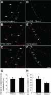
β-Catenin localization is not affected in scribble RNAi-expressing cells. (A–F) Confocal images of 10 DIV hippocampal neurons transfected with Syn-GFP and RNAi-C (A–C) or RNAi-3 (D–F) using Lipofectamine 2000 (<1% transfection efficiency) and immunostained for β-catenin. Syn-GFP puncta colocalize with β-catenin puncta in cells expressing RNAi-C (B and C; arrows). Neurons expressing RNAi-3 exhibit a diffuse pattern of Syn-GFP (D); however, β-catenin expression remains punctate (E and F; arrows). The average density (G) and size (H) of β-catenin puncta ± SE is similar in control and RNAi-expressing cells. N > 10 cells and >250 puncta from three cultures. p > 0.05 using Student's t test. Scale bar, 10 μm.
Figure 7.
Figure 7.
Scribble is diffusely distributed long the axon in cells lacking β-catenin. (A–H) Hippocampal neurons cultured from 10 DIV B6.129-Ctnnb1tm2Kem/KnwJ (homozygous β-catenin flox) mice were cotransfected using Lipofectamine 2000 (<1% transfection efficiency) with Syn-GFP and RFP-hScrib (A–D) or Syn-GFP, RFP-hScrib, and a construct expressing the Cre recombinase to ablate β-catenin (E–H). Confocal images demonstrate that Syn-GFP and RFP-hScrib are colocalized and are apposed to postsynaptic PSD-95 at synapses in control cells (C and D; arrows). Neurons expressing the Cre recombinase exhibit a diffuse expression of Syn-GFP and RFP-hScrib (E and F), whereas PSD-95 expression remains punctate (G and H; arrows). Immunopositive PSD-95 puncta that do not colocalize with Syn-GFP may represent immunostaining on neurons that are not transfected. (I–N) Confocal images of 10 DIV hippocampal neurons transfected with Syn-GFP and RFP-hScrib plus either β-cat FL (I–K) or β-catΔPDZb (L–N). In β-cat FL–expressing neurons, Syn-GFP puncta largely colocalize with RFP-hScrib puncta (I–K; arrows), whereas cells expressing β-catΔPDZb exhibit a diffuse pattern of Syn-GFP and RFP-hScrib expression (L–N). The normalized average coverage of RFP-hScrib along the axon ± SE is greater in cells lacking β-catenin (Cre+ cells) (O) and in cells expressing β-catΔPDZb (P), indicating a more diffuse distribution along the axon. N > 7 cells and >500 puncta from three different cultures. *p < 0.05, ***p < 0.0001 using Student's t test. Scale bars, 10 μm.

Similar articles

Cited by

References

    1. Ahmari S. E., Buchanan J., Smith S. J. Assembly of presynaptic active zones from cytoplasmic transport packets. Nat. Neurosci. 2000;3:445–451. - PubMed
    1. Audebert S., et al. Mammalian Scribble forms a tight complex with the betaPIX exchange factor. Curr. Biol. 2004;14:987–995. - PubMed
    1. Bamji S. X., Rico B., Kimes N., Reichardt L. F. BDNF mobilizes synaptic vesicles and enhances synapse formation by disrupting cadherin-beta-catenin interactions. J. Cell Biol. 2006;174:289–299. - PMC - PubMed
    1. Bamji S. X., Shimazu K., Kimes N., Huelsken J., Birchmeier W., Lu B., Reichardt L. F. Role of beta-catenin in synaptic vesicle localization and presynaptic assembly. Neuron. 2003;40:719–731. - PMC - PubMed
    1. Bozdagi O., Shan W., Tanaka H., Benson D. L., Huntley G. W. Increasing numbers of synaptic puncta during late-phase LTP: N-cadherin is synthesized, recruited to synaptic sites, and required for potentiation. Neuron. 2000;28:245–259. - PubMed

Publication types

Substances