. 2009 Jul;20(14):3330-41.
doi: 10.1091/mbc.e08-09-0953. Epub 2009 May 20.

Dislocation of HMG-CoA reductase and Insig-1, two polytopic endoplasmic reticulum proteins, en route to proteasomal degradation

Affiliations

Dislocation of HMG-CoA reductase and Insig-1, two polytopic endoplasmic reticulum proteins, en route to proteasomal degradation

Gil S Leichner et al. Mol Biol Cell. 2009 Jul.

Abstract

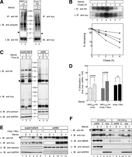
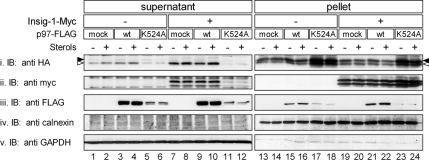
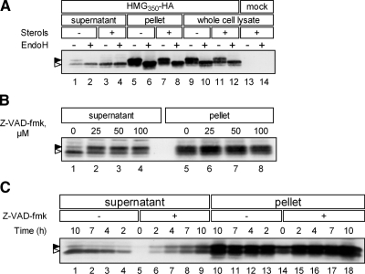
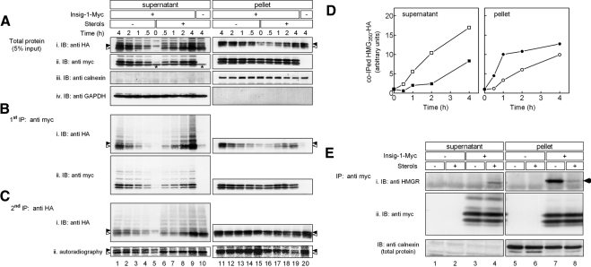
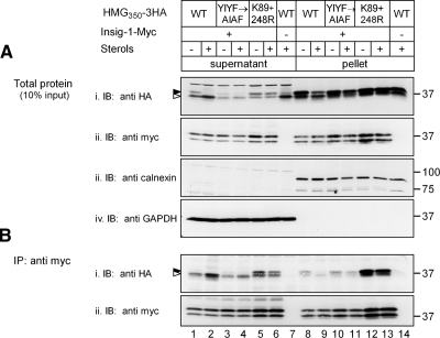
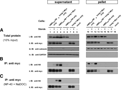
The endoplasmic reticulum (ER) glycoprotein HMG-CoA reductase (HMGR) catalyzes the rate-limiting step in sterols biosynthesis. Mammalian HMGR is ubiquitinated and degraded by the proteasome when sterols accumulate in cells, representing the best example for metabolically controlled ER-associated degradation (ERAD). This regulated degradation involves the short-lived ER protein Insig-1. Here, we investigated the dislocation of these ERAD substrates to the cytosol en route to proteasomal degradation. We show that the tagged HMGR membrane region, HMG(350)-HA, the endogenous HMGR, and Insig-1-Myc, all polytopic membrane proteins, dislocate to the cytosol as intact full-length polypeptides. Dislocation of HMG(350)-HA and Insig-1-Myc requires metabolic energy and involves the AAA-ATPase p97/VCP. Sterols stimulate HMG(350)-HA and HMGR release to the cytosol concurrent with removal of their N-glycan by cytosolic peptide:N-glycanase. Sterols neither accelerate dislocation nor stimulate deglycosylation of ubiquitination-defective HMG(350)-HA((K89 + 248R)) mutant. Dislocation of HMG(350)-HA depends on Insig-1-Myc, whose dislocation and degradation are sterol independent. Coimmunoprecipitation experiments demonstrate sterol-stimulated association between HMG(350)-HA and Insig-1-Myc. Sterols do not enhance binding to Insig-1-Myc of HMG(350)-HA mutated in its sterol-sensing domain or of HMG(350)-HA((K89 + 248R)). Wild-type HMG(350)-HA and Insig-1-Myc coimmunoprecipitate from the soluble fraction only when both proteins were coexpressed in the same cell, indicating their encounter before or during dislocation, raising the possibility that they are dislocated as a tightly bound complex.


Figures

Figure 1.
Figure 1.
Ubiquitination, degradation, and dislocation of HMG350-HA, HMGR and Insig-1-Myc. (A) Sterol-depleted cells, stably expressing either HMG350-HA or Insig-1-Myc, were incubated for 2 h with ALLN (65 μM) with (+) or without (−) sterols (2 μg/ml 25-hydroxycholesterol plus 20 μg/ml cholesterol). Cells were lysed in solution D, and proteins from the postnuclear supernatant were immunoprecipitated (IP) with rabbit polyclonal anti-HA (lanes 1 and 2) or anti-myc (lanes 3 and 4) antibodies. The immune complexes were resolved by 5–15% SDS-PAGE, electroblotted onto nitrocellulose, and the membrane was first probed (IB) with a mouse anti-ubiquitin monoclonal antibody (top) and then reprobed with mouse anti-HA and anti-myc monoclonal antibodies (bottom). Arrowheads indicate the migration of the immunoprecipitated proteins. Mr markers (in kilodaltons) are shown. (B) Sterol-depleted cells, as in A, were pulse labeled for 30 min with 35S-protein label mix (100 μCi/dish; 300 μCi/ml) and chased for the indicated time in unlabeled medium in the absence or presence of sterols. Cells were lysed in solution D, and proteins were immunoprecipitated with the mouse monoclonal anti-HA (top) or anti-myc (bottom) antibodies. The immune complexes were resolved by SDS-PAGE followed by fluorography. Densitometric analysis of the gels is shown. •○, HMG350-HA; ■□, Insig-1-Myc; closed symbols, sterol-depleted cells; open symbols, sterol-treated cells. (C) Cells expressing both HMG350-HA and Insig-1-Myc, were incubated for the indicated time with ALLN with (+) or without (−) sterols, as described in A. Cells were permeabilized in solution C and fractionated by centrifugation, as described under Materials and Methods. Aliquots of the supernatant (60 μg of protein) and solubilized membranes (20 μg of protein) were IB and reprobed with the indicated antibodies. Closed and open arrowheads indicate glycosylated and un/deglycosylated forms of HMG350-HA, respectively. The asterisk (*) denotes a protein in the supernatant fraction that cross-reacted with the anti-myc antibody. Mr markers (in kilodaltons) are shown. (D) Densitometric analysis of experiments, as in C, of cells expressing only HMG350-HA (light gray bars; n = 12), or cells expressing both HMG350-HA and Insig-1-Myc or Insig-1-Myc alone probed with anti-HA (dark gray bars; n = 11) or with anti-Myc (black bars; n = 8) antibodies. The results (mean ± SEM) were evaluated by Student's t test, and p values are indicated; n.s., not significant. (E) On day 1, SRD-15 cells were transfected with 2 μg of pIRESneo2-HMG350-HA along with 0.5 μg of an empty vector (−) or pcDNA3.1-Insig-1-Myc (+), as indicated. On day 2, the cells were replated in triplicate dishes in medium lacking sterols but containing 50 μM lovastatin and 50 μM MVA. On day 3, the cells were harvested immediately (0) or incubated for 2 h with 65 μM ALLN in the presence (+) or absence (−) of sterols. Cells were permeabilized, and supernatant (45 μg of protein) and pellet (22 μg of protein) fractions were analyzed by immunoblotting with the indicated antibodies. Asterisks denote a protein in the supernatant fraction that cross-reacted with the anti-myc antibody. (F) Sterol-depleted cells expressing both HMG350-HA and Insig-1-Myc, were harvested immediately (0) or incubated for 2 h with 65 μM ALLN with (+) or without (−) sterols. Cells were permeabilized and fractionated by centrifugation, as described under Materials and Methods. The 20,000 × g supernatant was further centrifuged at 100,000 × g for 30 min. Samples of the pellet (100 μg of protein) and supernatant (30 μg of protein) fractions were analyzed by SDS-PAGE and IB with the indicated antibodies. Closed and open arrowheads indicate glycosylated and un/deglycosylated forms of HMG350-HA, respectively. The asterisk (*) denotes a protein in the supernatant fraction that cross-reacted with the anti-myc antibody.
Figure 2.
Figure 2.
Dislocation of HMG350-HA and Insig-1-Myc involves p97/VCP. On day 1, HEK293 cells were transfected with a plasmid mixture consisting of 3 μg of pIRESneo2-HMG350-HA, 0.5 μg of pcDNA3.1-Insig-1-Myc, and 0.5 μg of wild-type or K524A mutant of pcDNA3.1-p97-FLAG, as indicated. Total plasmid concentration was adjusted to 4 μg/dish with empty pcDNA3 vector. Forty-eight hours later, the cells were switched to medium B for additional 16 h after which they were treated for 2 h with ALLN (65 μM) with (+) or without (−) sterols. Cells were permeabilized, and supernatant (60 μg of protein) and pellet (20 μg of protein) fractions were analyzed by immunoblotting with the indicated antibodies. Closed and open arrowheads indicate glycosylated and un/deglycosylated forms of HMG350-HA, respectively.
Figure 3.
Figure 3.
Cytosolic PNGase removes N-glycan from dislocated HMG350-HA. (A) Mock-transfected cells or cells expressing HMG350-HA were incubated for 2 h with ALLN (65 μM) with (+) or without (−) sterols. Cells were permeabilized in solution C and fractionated (lanes 1–8), or solubilized in solution D (lanes 9–14). Aliquots of supernatant (75 μg of protein), solubilized pellet (20 μg of protein), or whole cell lysates (120 μg protein) were treated (+) or mock treated (−) with 1000 U of EndoH and analyzed. (B) Cells were preincubated for 1 h with the indicated concentrations of Z-VAD-fmk before addition of ALLN and sterols. The cells were incubated for additional 2 h and then permeabilized and fractionated. Aliquots of supernatant (60 μg of protein) and solubilized pellet (20 μg of protein) were analyzed. (C) Cells were incubated with ALLN and sterols, in the absence (−) or presence (+) of Z-VAD-fmk (50 μM). At the indicated time points, cells were permeabilized and fractionated and aliquots of supernatant (75 μg of protein) and solubilized pellet (20 μg of protein) were analyzed. All samples were resolved by 5–15% SDS-PAGE and immunoblotted with anti-HA monoclonal antibody. Closed and open arrowheads indicate glycosylated and un/deglycosylated forms of HMG350-HA, respectively.
Figure 4.
Figure 4.
Sterol-stimulated formation and dislocation of HMGR · Insig-1-Myc complex. Sterol-depleted cells that express HMG350-HA together (+) or without (−) Insig-1-Myc were pulse-labeled for 30min with of 35S–cell-labeling reagent (100 μCi). Cells were chased in the presence of ALLN with (+) or without (−) sterols, permeabilized, and fractionated. (A) Aliquots of supernatant and solubilized pellet (5% of samples) were analyzed by SDS-PAGE and immunoblotting (IB) with the indicated antibodies. (B) The rest 95% of the samples were first subjected to immunoprecipitation (first IP) of Insig-1-Myc with a mouse monoclonal anti-myc antibody, and immune complexes were analyzed by SDS-PAGE and IB with rabbit antibodies for the presence of Insig-1-Myc (ii) and coprecipitated HMG350-HA (i). (C) The supernatants from the first IP were cleared with protein A-Sepharose beads and reprecipitated (second IP) with a mouse monoclonal anti-HA antibody and immune complexes were analyzed by SDS-PAGE for the presence of HMG350-HA by IB with a rabbit anti-HA antibody (i) and by direct autoradiography (ii). Closed and open arrowheads indicate glycosylated and un/deglycosylated forms of HMG350-HA, respectively. Asterisks (Aii, lanes 5 and 10) denote a protein in the supernatant fraction that cross-reacted with the anti-myc antibody. (D) Densitometric analysis of the blots. The HA immunoreactive signal in Bi (including trailing smear) was divided by the Myc-immunoreactive signal in Bii (including trailing smear), setting the ratio at time zero as one. Results are the mean of two independent experiments. Closed symbols, sterol-depleted cells; open symbols, sterol-treated cells. (E) Sterol-depleted naïve or Insig-1-Myc–expressing cells were incubated for 2 h with ALLN and sterols, as indicated. Cells were permeabilized in solution C and fractionated. Proteins from the supernatant and pellet fractions were IP with rabbit polyclonal anti-myc antibodies. The immune complexes were resolved and IB with a mouse anti-HMGR monoclonal antibody (i; arrow) and then reprobed with mouse anti-myc monoclonal antibodies (ii). Aliquots of the fractions, representing 5% of protein input, were immunoblotted with rabbit anti-calnexin antibody.
Figure 5.
Figure 5.
Interactions with Insig-1-Myc and dislocation of HMG350-HA mutants. Cells expressing the indicated constructs of HMG350-HA, together (+) or without (−) Insig-1-Myc, were incubated for 2h with ALLN with (+) or without (−) sterols, permeabilized in solution C and fractionated. (A) Aliquots of supernatant and solubilized pellet (10% of samples) were analyzed by SDS-PAGE and IB with the indicated antibodies. (B) The rest 90% of the samples were subjected to IP with an anti-myc monoclonal antibody, and immune complexes were analyzed by SDS-PAGE and immunoblotting (IB) with rabbit anti-myc and anti-HA antibodies for the presence of Insig-1-Myc (ii) and coprecipitated HMG350-HA (i), respectively. Closed and open arrowheads indicate glycosylated and un/deglycosylated forms of HMG350-HA, respectively. Mr markers (in kilodaltons) are shown.
Figure 6.
Figure 6.
Interaction between HMG350-HA and Insig-1-Myc requires their expression in the same cell. Sterol-depleted cells expressing either HMG350-HA, Insig-1-Myc, or both, and a mixture of cells expressing either proteins, were incubated for 2h with ALLN with (+) or without (−) sterols, and cells were permeabilized and fractionated. (A) Aliquots of supernatant (5%) and solubilized pellet (10%) (see Note) were analyzed by SDS-PAGE and immunoblotting (IB) with the indicated antibodies. The rest 95% of the supernatant fraction was divided into two equal portions. (B) One portion was directly immunoprecipitated (IP) with an anti-myc monoclonal antibody and immune complexes were analyzed by SDS-PAGE and immunoblotting with rabbit anti-myc and anti-HA antibodies, for the presence of Insig-1-Myc (ii) and coprecipitated HMG350-HA (i), respectively. (C) To the other supernatant portion, NP-40 and NaDOC were added to 1% each, and the samples were immunoprecipitated and immunoblotted as described in B. Note: Because the supernatant was divided into two portions, the 5% sample of the initial fraction actually represents 10% of total protein input in each portion used for the analyses in B and C.

Similar articles

Cited by

References

    1. Afshar N., Black B. E., Paschal B. M. Retrotranslocation of the chaperone calreticulin from the endoplasmic reticulum lumen to the cytosol. Mol. Cell Biol. 2005;25:8844–8853. - PMC - PubMed
    1. Baker B. M., Tortorella D. Dislocation of an ER membrane glycoprotein involves the formation of partially dislocated ubiquitinated polypeptides. J. Biol. Chem. 2007;282:26845–26856. - PubMed
    1. Bar-Nun S. The role of p97/Cdc48p in endoplasmic reticulum-associated degradation: from the immune system to yeast. Curr. Top. Microbiol. Immunol. 2005;300:95–125. - PubMed
    1. Brodsky J. L. The protective and destructive roles played by molecular chaperones during ERAD (endoplasmic-reticulum-associated degradation) Biochem. J. 2007;404:353–363. - PMC - PubMed
    1. Brown D. A., Simoni R. D. Biogenesis of 3-hydroxy-3-methylglutaryl-coenzyme A reductase, an integral glycoprotein of the endoplasmic reticulum. Proc. Natl. Acad. Sci. USA. 1984;81:1674–1678. - PMC - PubMed

MeSH terms