. 2009 May 20;29(20):6436-48.
doi: 10.1523/JNEUROSCI.5479-08.2009.

Context-specific grasp movement representation in the macaque anterior intraparietal area

Affiliations

Context-specific grasp movement representation in the macaque anterior intraparietal area

Markus A Baumann et al. J Neurosci. .

Abstract

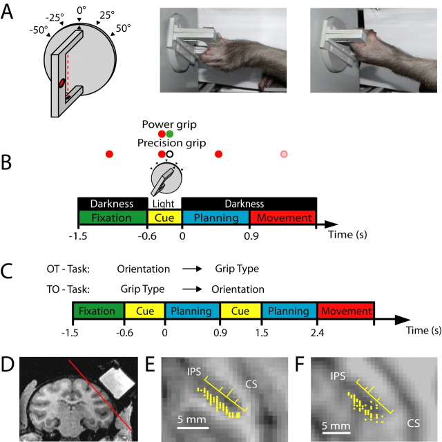
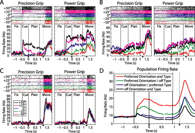
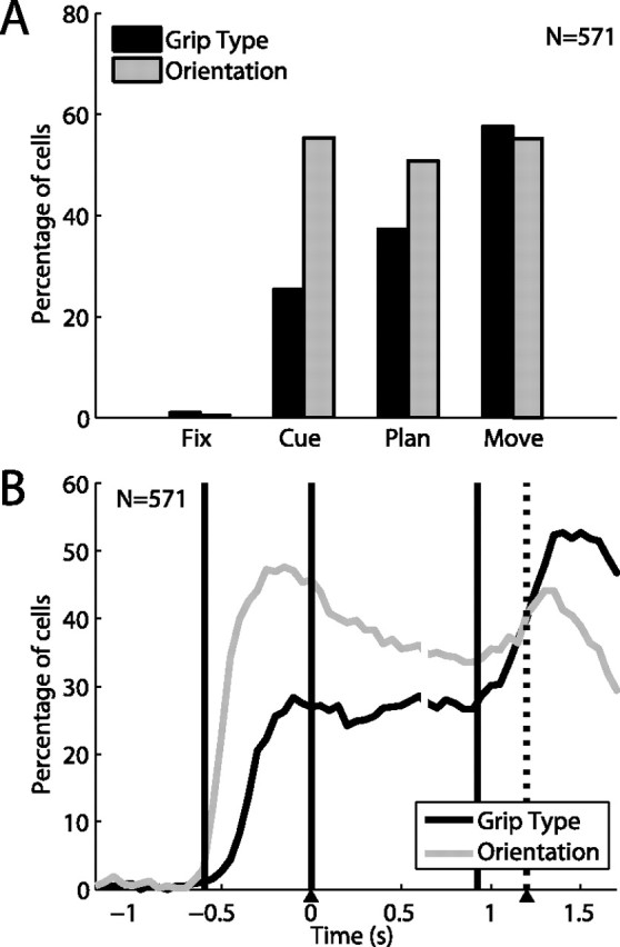
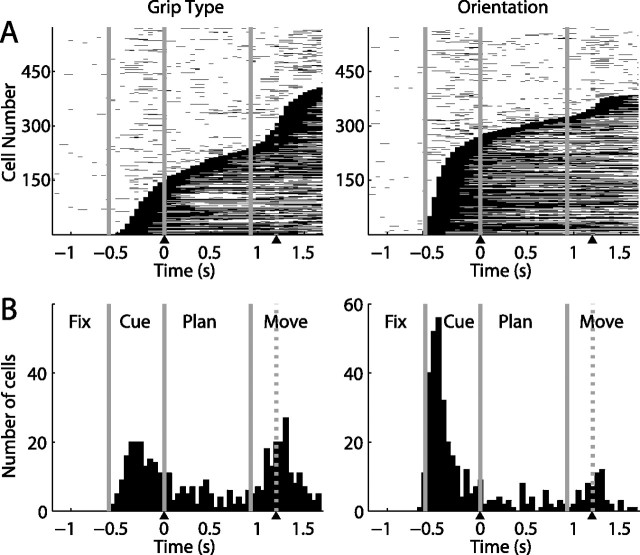
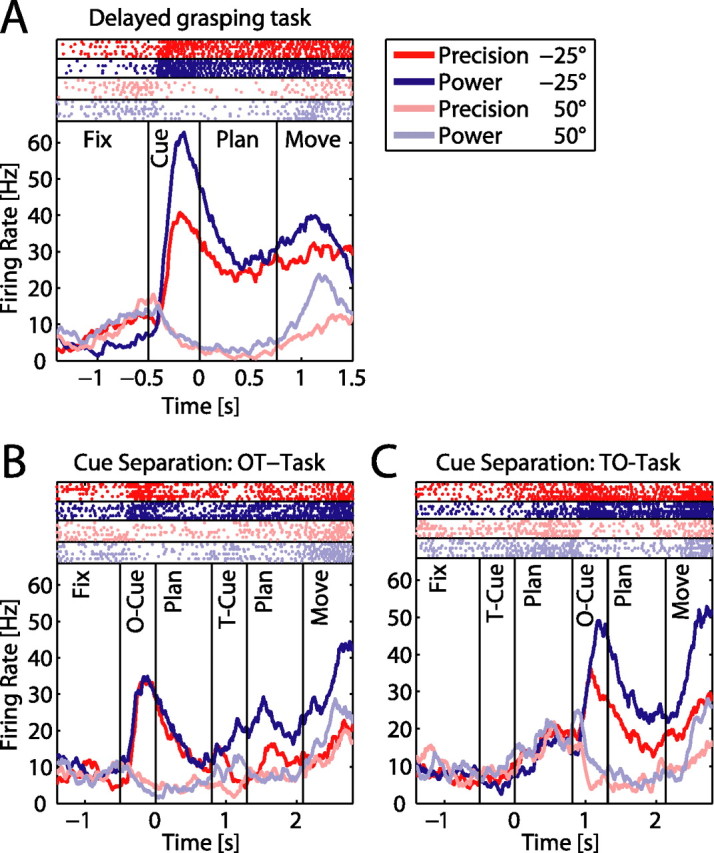
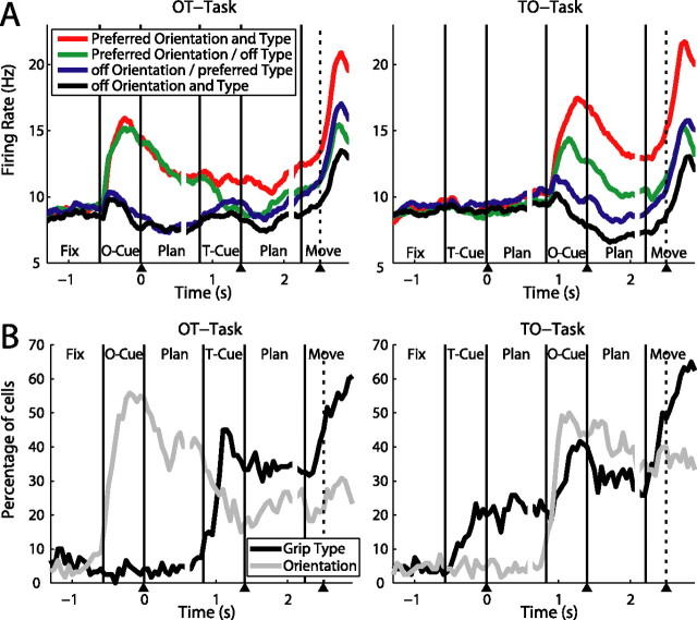
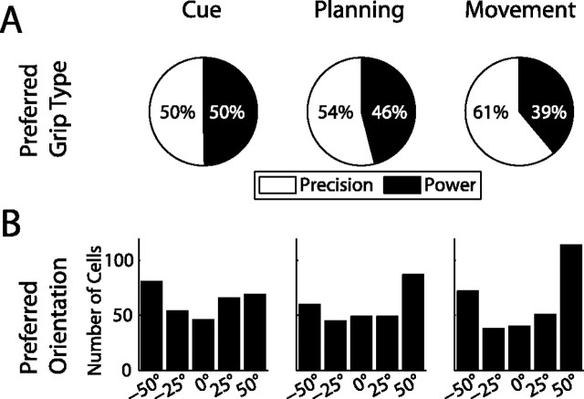
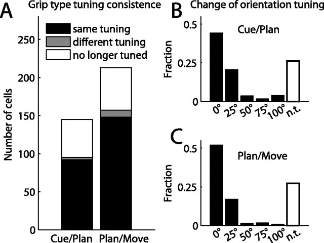
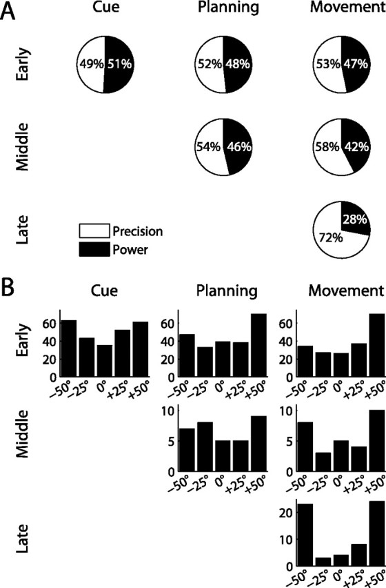
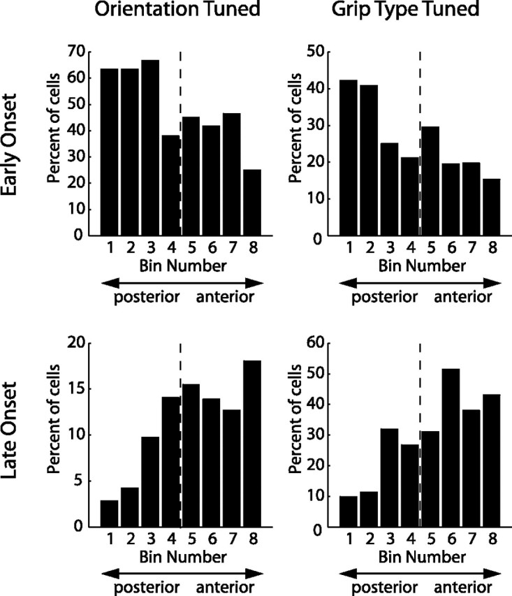
To perform grasping movements, the hand is shaped according to the form of the target object and the intended manipulation, which in turn depends on the context of the action. The anterior intraparietal cortex (AIP) is strongly involved in the sensorimotor transformation of grasping movements, but the extent to which it encodes context-specific information for hand grasping is unclear. To explore this issue, we recorded 571 single-units in AIP of two macaques during a delayed grasping task, in which animals were instructed by an external context cue (LED) to perform power or precision grips on a handle that was presented in various orientations. While 55% of the recorded neurons encoded the object orientation from the cue epoch on, the number of cells encoding the grip type increased from 25% during the cue epoch to 58% during movement execution. Furthermore, a classification of cells according to the time of their tuning onset revealed differences in the function and anatomical location of early- versus late-tuned cells. In a cue separation task, when the object was presented first, neurons representing power or precision grips were activated simultaneously until the actual grip type was instructed. In contrast, when the grasp type instruction was presented before the object, type information was only weakly represented in AIP, but was strongly encoded after the grasp target was revealed. We conclude that AIP encodes context specific hand grasping movements to perceived objects, but in the absence of a grasp target, the encoding of context information is weak.


Figures

Figure 1.
Figure 1.
Task paradigm and recording penetrations. A, Sketch of the handle (left) and photographs of a monkey performing a precision grip (middle) and a power grip (right). In the drawing, the red dotted line indicates a light barrier for detecting power grips, and the red oval indicates a touch sensor in a groove for sensing precision grips (a second sensor is located on the opposite side of the handle). The handle was presented in five different orientations. B, Delayed grasping task. Trials were divided into four epochs: fixation, cue, planning, and movement. Monkeys initiated trials by placing both hands on rest sensors and fixating a red LED in the dark. After a variable delay (fixation, 700–1100 ms), the handle was illuminated for 600 ms (cue), revealing its orientation. At the same time, a second colored LED (“context cue”) was illuminated, which instructed the animal about the required grip type (power or precision). After a variable delay (planning, 700–1100 ms), the dimming of the fixation light served as the go signal to initiate movement execution. All trial conditions were randomly interleaved. C, Cue separation task. Modified task from B, with the cues for grip type and orientation presented consecutively and with each cue followed by a separate planning period. In one version of this task (OT task), the orientation information preceded the grip type information, while in the other version the cue sequence was reversed (TO task). D, Coronal MRI section (monkey J) with the recording chamber on the right hemisphere filled with contrast medium. The red line indicates the position of the oblique section in E. E, F, Maps of recording electrode penetrations (yellow dots) in monkeys J and L, respectively. The yellow ruler indicates the median (long tick mark) and quartiles of the recording distribution along the intraparietal sulcus (IPS). CS, Central sulcus.
Figure 2.
Figure 2.
Three example neurons with different tuning onsets. For each neuron, precision grip trials are shown on the left panel and power grips on the right panel. Different colors indicate various handle orientations, for which spike rasters (on top) and averaged firing rates (at bottom) are shown individually. The dotted line within the movement epoch indicates the release of the hand rest button (movement start). All trials are aligned to both the end of the cue epoch and the start of the movement (arrow heads below); gaps in the curves (at ∼0.6 s) indicate the realignment. A, Neuron that exhibits tuning for the handle orientation and the instructed grip type starting in the cue period and extending until movement execution. B, Neuron with tuning for the handle orientation starting in cue, but with significant grip type modulation only during movement execution. C, Neuron showing no response during cue presentation and movement planning, but with a strong selectivity for precision grips during movement execution without significant orientation tuning. D, Population firing rate across all 571 neurons for each combination of the cells' preferred and nonpreferred grip type and orientation.
Figure 3.
Figure 3.
Orientation and grip type tuning in the neuronal population (n = 571). A, Fraction of cells showing tuning for grip type (black) and handle orientation (gray) in the different task epochs (two-way ANOVA; see Materials and Methods). Tuning for object orientation was constant from cue to movement, while grip type tuning increased over time. B, Percentage of tuned cells in a sliding window (width: 200 ms, centered on each data point). During cue, tuning for orientation started ∼150 ms earlier than for grip type. Grip type tuning increased in two steps: one during cue and one during the movement epoch. Trials are aligned on cue offset and movement onset (as in Fig. 2).
Figure 4.
Figure 4.
Times with significant tuning in the neuronal population. A, Sliding window analysis (two-way ANOVA) for each cell (y-axis) at each time step (x-axis). Significant grip type (left) and orientation tuning (right) is indicated by black squares (p < 0.01). Cells are ordered by tuning onset (first occurrence of five consecutive significant steps). B, Histogram of tuning onset for grip type (left) and orientation (right) across the population.
Figure 5.
Figure 5.
Neural activity in the cue separation task. Panels show one example neuron during the delayed grasping task (A) and the cue separation task: OT task (B), TO task (C). Different grip types are shown in red (precision) and blue (power), while the two grip orientations are shown in light and dark color. A, The neuron was early tuned for both parameters, showing highest activity for power grips at −25° orientation. B, In the OT task, presentation of the object in the −25° orientation evoked a strong response, which was then differentially modulated for the two grip types after the second cue. C, In the TO task, the cell did not respond to the type cue when presented in the absence of the object. However, the cell responded vigorously after the orientation cue with a preference for power grips in the preferred orientation.
Figure 6.
Figure 6.
Population analysis of the cue separation task. A, Population firing rates in the cue separation task (N = 120) with OT task on the left and TO task on the right panel. For each cell, its preferred type and orientation was established in the delayed grasping task (data not shown). B, Fraction of cells that were significantly tuned by grip type and orientation in the course of the OT and TO task (sliding window ANOVA as in Fig. 3B).
Figure 7.
Figure 7.
Distribution of preferred grip type and orientation in various task epochs. A, Ratio of cells preferring precision (white) or power grip (black). From cue to movement, the fraction of cells encoding precision grip increased substantially. B, Number of cells preferring each of the five orientations. In the movement epoch, the distribution shifts in favor of terminal orientations (±50°).
Figure 8.
Figure 8.
Tuning consistency across task epochs. A, Grip type tuning. Histogram bars indicate the number of cells that stay tuned for the same grip (black), change preference to the opposite grip (gray), or lose their tuning (white) when transitioning between consecutive task epochs (cue–planning and planning–movement). B, C, Change of orientation tuning in consecutive task epochs: cue–planning (B) and planning–movement (C). Histograms show the fraction of cells for which the preferred orientations in the two epochs were the same (0° shift), neighboring orientations (25°), or further apart (50–100°), and of cells that lost their tuning (white bars). Preferred orientation shifts of >25° were rare. In general, cells were tuned consistently over time.
Figure 9.
Figure 9.
Distribution of preferred grip type and orientation in different cell classes. A, Ratio of precision and power preference in cell groups with early (top row), intermediate (middle), and late (bottom) tuning onset for grip type. In all three task epochs, early-tuned cells preferred power grips and precision grips approximately equally often. In contrast, ∼70% of late tuning cells preferred precision grips. B, Number of cells with a particular orientation preference for the three cell classes. In early orientation-tuned cells, the portion of cells that preferred extreme orientations (±50°) changed little from cue (49%) to movement (53%), while 78% of cells with a late onset of orientation tuning preferred extreme orientations.
Figure 10.
Figure 10.
Anatomical distribution of different cell classes. Cells (N = 571) were distributed into eight bins according to their location along the intraparietal sulcus, such that each bin contained the same number of cells. Bin 1 contained the most posterior and bin 8 the most anterior cells (x-axis). Individual panels show the distribution along the intraparietal sulcus (IPS) for a particular cell class (left: orientation tuned, right: grip type tuned, top: early onset, bottom: late onset). Histograms display the fraction of cells in each bin that belonged to the respective cell class. Early onset cells showed a decreasing, late onset cells an increasing gradient from posterior to anterior. Dashed line, Median of the population.

Comment in

References

    1. Allport DA. Selection for action: some behavioral and neurophysiological considerations of attention and action. In: Heuer H, Sanders AF, editors. Perspectives on perception and action. Hillsdale, NJ: Erlbaum; 1987. pp. 395–419.
    1. Amemori K, Sawaguchi T. Rule-dependent shifting of sensorimotor representation in the primate prefrontal cortex. Eur J Neurosci. 2006;23:1895–1909. - PubMed
    1. Andersen RA. Multimodal integration for the representation of space in the posterior parietal cortex. Philos Trans R Soc Lond B Biol Sci. 1997;352:1421–1428. - PMC - PubMed
    1. Andersen RA, Buneo CA. Intentional maps in posterior parietal cortex. Annu Rev Neurosci. 2002;25:189–220. - PubMed
    1. Balint R. Seelenlähmung des “Schauens”, optische Ataxie, räumliche Störung der Aufmerksamkeit. Monatsschr Psychiatr Neurol. 1909;25:51–81.

Publication types

LinkOut - more resources