. 2009 May 20;29(20):6568-79.
doi: 10.1523/JNEUROSCI.0181-09.2009.

Mobilization of calcium from intracellular stores facilitates somatodendritic dopamine release

Affiliations

Mobilization of calcium from intracellular stores facilitates somatodendritic dopamine release

Jyoti C Patel et al. J Neurosci. .

Abstract

Somatodendritic dopamine (DA) release in the substantia nigra pars compacta (SNc) shows a limited dependence on extracellular calcium concentration ([Ca(2+)](o)), suggesting the involvement of intracellular Ca(2+) stores. Here, using immunocytochemistry we demonstrate the presence of the sarcoplasmic/endoplasmic reticulum Ca(2+)-ATPase 2 (SERCA2) that sequesters cytosolic Ca(2+) into the endoplasmic reticulum (ER), as well as inositol 1,4,5-triphosphate receptors (IP(3)Rs) and ryanodine receptors (RyRs) in DAergic neurons. Notably, RyRs were clustered at the plasma membrane, poised for activation by Ca(2+) entry. Using fast-scan cyclic voltammetry to monitor evoked extracellular DA concentration ([DA](o)) in midbrain slices, we found that SERCA inhibition by cyclopiazonic acid (CPA) decreased evoked [DA](o) in the SNc, indicating a functional role for ER Ca(2+) stores in somatodendritic DA release. Implicating IP(3)R-dependent stores, an IP(3)R antagonist, 2-APB, also decreased evoked [DA](o). Moreover, DHPG, an agonist of group I metabotropic glutamate receptors (mGluR1s, which couple to IP(3) production), increased somatodendritic DA release, whereas CPCCOEt, an mGluR1 antagonist, suppressed it. Release suppression by mGluR1 blockade was prevented by 2-APB or CPA, indicating facilitation of DA release by endogenous glutamate acting via mGluR1s and IP(3)R-gated Ca(2+) stores. Similarly, activation of RyRs by caffeine increased [Ca(2+)](i) and elevated evoked [DA](o). The increase in DA release was prevented by a RyR blocker, dantrolene, and by CPA. Importantly, the efficacy of dantrolene was enhanced in low [Ca(2+)](o), suggesting a mechanism for maintenance of somatodendritic DA release with limited Ca(2+) entry. Thus, both mGluR1-linked IP(3)R- and RyR-dependent ER Ca(2+) stores facilitate somatodendritic DA release in the SNc.


Figures
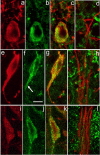
Figure 1.
Figure 1.
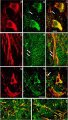
ER Ca2+ stores protein immunoreactivity in nigral DAergic neurons. a–c, SERCA2 immunoreactivity in DAergic neurons in the SNc. Immunostaining for TH (red) (a) and SERCA2 (green) (b) with merged images for TH and SERCA (overlap appears yellow) (c). In the perikaryal cytoplasm, SERCA2 immunoreactivity is observed as a ring around the nucleus that extends almost to the perikaryal edge. Additional SERCA staining is seen within the most proximal portion of TH-ir dendrites (TH-ir profiles above perikaryon in c). d, However, SERCA staining is absent from DAergic dendrites within SNr. e–g, Localization of IP3Rs in DAergic neurons in the SNc. Immunostaining for TH (red) (e) and IP3R (green) (f) with merged images for TH and IP3R (g). Note that immunoreactivity to IP3R and TH colocalizes in the perikaryon and extends down a proximal dendrite (f, arrow). h, In SNr, however, IP3R immunoreactivity (green) does not colocalize with that of TH (red), implying minimal IP3R expression in distal DAergic dendrites. i–k, Localization of RyRs in DAergic neurons in the SNc. Immunostaining for TH (red) (i) and RyR (green) (j) with merged images for TH and RyR (k). Note that RyR immunostaining includes a mixture of large puncta located at the edge of the perikaryon and smaller puncta located in the perikaryal cytoplasm. I, RyR immunoreactivity (green) does not colocalize with that of TH within SNr, implying low levels in distal dendrites. Scale bar: (in f) a–I, 10 μm.
Figure 2.
Figure 2.
mGluR1α and Cav1.3 immunoreactivity in nigral DAergic neurons. a–c, Immunostaining for TH (red) (a) and mGluR1α (green) (b) with merged images for TH and mGluR1α (overlap appears yellow) (c). The mGluR1α immunoreactivity appears as puncta that colocalize with TH in the perikarya and primary dendrites of DAergic neurons in SNc. Arrows in b and c indicate corresponding locations at which colocalization of mGluR1α immunoreactivity and TH immunoreactivity occurs in dendritic processes within SNc. d–f, A moderate level of mGluR1α staining is seen within TH-ir dendrites in SNr. Paired arrows in e and f point to colocalization of mGluR1α and TH immunoreactivity in a dendritic profile. g–i, Localization of Cav1.3, an L-type Ca2+ channel subunit, in DAergic neuronal perikarya in the SNc. Immunostaining for TH (red) (g) and Cav1.3 (green) (h) with merged images for TH and Cav1.3 (i). Cav1.3 immunoreactivity is punctate. j, k, Colocalization of immunoreactivity to TH and Cav1.3 in DAergic processes at the border of SNc/SNr (j) and deep within SNr (k). Scale bar: (in c) a–f, 10 μm; (in h) g–k, 20 μm.
Figure 3.
Figure 3.
Ca2+ entry and intracellular Ca2+ facilitate somatodendritic DA release. a, Average [DA]o versus time profiles evoked by local stimulation (30 pulses, 10 Hz) in the SNc in the absence and presence of a nonselective Ca2+ channel blocker, Cd2+ (100 μm, n = 6). b, Average [DA]o versus time profiles in SNc in the absence and presence of a fast-acting Ca2+ chelator BAPTA-AM (BAPTA) (50 μm, n = 6). c, Summary of the effect of Cd2+ and BAPTA on peak [DA]o; evoked [DA]o was measured at the time point of control peak [DA]o, which was taken as 100%. Blockade of stimulus-induced Ca2+ entry by Cd2+ abolished evoked [DA]o (n = 6, ***p < 0.001 vs control), confirming that Ca2+ entry is required to trigger evoked somatodendritic DA release. Buffering of stimulus-induced intracellular Ca2+ by BAPTA decreased evoked [DA]o (n = 6, **p < 0.01 vs control), demonstrating the involvement of intracellular Ca2+ elevation in evoked somatodendritic DA release.
Figure 4.
Figure 4.
Effect of SERCA inhibition on somatodendritic DA release. a, Average [DA]o versus time profiles in the SNc evoked by local stimulation (30 pulses, 10 Hz) in the absence and presence of a membrane-permeable SERCA inhibitor, CPA (30 μm, n = 6). b, Summary of the effect of CPA on peak [DA]o; control peak [DA]o was taken as 100%. Inhibition of SERCA by CPA decreased evoked [DA]o (n = 6, **p < 0.01 vs control), demonstrating the involvement of ER Ca2+ stores in somatodendritic DA release.
Figure 5.
Figure 5.
Regulation of somatodendritic DA release by mGluR1 activation of IP3R-gated intracellular Ca2+ stores. a, Average [DA]o versus time profiles in SNc evoked by local stimulation (30 pulses, 10 Hz) in the absence and presence of a membrane-permeable IP3R inhibitor, 2-APB (left) (100 μm, n = 8). b, Summary of the effect of 2-APB on peak [DA]o; control peak [DA]o (Con) was taken as 100%. Inhibition of IP3Rs by 2-APB decreased evoked [DA]o (n = 8, ***p < 0.001 vs control), indicating involvement of Ca2+ mobilization from IP3R-gated stores in somatodendritic DA release. c, Average [DA]o versus time profiles in SNc in the absence and presence of an mGluR1 agonist DHPG (left) (1 μm, n = 8) or the mGluR1 antagonist CPCCOEt (right) (100 μm, n = 9). d, Summary of the effect of DHPG and CPCCOEt (CPC) on peak [DA]o; control peak [DA]o was taken as 100%. Activation of the IP3R-dependent mGluR1 pathway by DHPG significantly increased evoked [DA]o (n = 8, *p < 0.05 vs control), implicating a role for mGluR1-gated Ca2+ stores in somatodendritic DA release. Blockade of mGluR1s with CPCCOEt decreased evoked [DA]o (n = 9, ***p < 0.001), indicating that endogenously released glutamate normally facilitates somatodendritic DA release via activation of mGluR1s. e, Average [DA]o versus time profiles in SNc with CPCCOEt (100 μm) after pretreatment with the IP3R antagonist 2-APB (left) (100 μm, n = 6) or the SERCA inhibitor CPA (right) (30 μm, n = 6). f, Summary of the effect of CPCCOEt in 2-APB or CPA on peak [DA]o; control peak [DA]o in either 2-APB or CPA alone was taken as 100%. Suppression of evoked [DA]oby CPCCOEt was prevented by pretreatment with 2-APB (n = 6, p > 0.05, CPC + 2-APB vs 2-APB alone) or CPA (n = 6, p > 0.05, CPC + CPA vs CPA alone), demonstrating that activation of mGluR1s by endogenously released glutamate involves mobilization of Ca2+ from IP3R-gated ER stores.
Figure 6.
Figure 6.
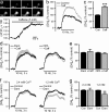
Regulation of somatodendritic DA release by RyR-gated intracellular Ca2+ stores. a, Representative record of Fluo-5F fluorescence during RyR-activation by caffeine (5 mm), showing the time course of the resultant increase in [Ca2+]i in an SNc DAergic neuron. Recordings were made in nominally zero [Ca2+]o and in the presence of TTX (1 μm). b, Average [DA]o versus time profiles in the SNc evoked by local stimulation (30 pulses, 10 Hz) in the absence and presence of caffeine (Caff) (5 mm, n = 6). c, Summary of the effect of caffeine on peak [DA]o; control peak [DA]o (Con) was taken as 100%. Activation of RyRs by caffeine increased evoked [DA]o (n = 6, ***p < 0.001 vs control), implicating RyR-gated Ca2+ stores in somatodendritic DA release. d, Average [DA]o versus time profiles in SNc with caffeine (5 mm) after pretreatment with dantrolene (Dant) (10 μm), a RyR blocker (left, n = 8) or CPA (30 μm), a SERCA inhibitor (right, n = 6). e, Summary of the effect of caffeine in dantrolene or CPA on peak [DA]o; control peak [DA]o in either dantrolene or CPA alone was taken as 100%. Enhancement of evoked [DA]o by caffeine is prevented by pretreatment with dantrolene (n = 8, p > 0.05, Caff + Dant vs Dant alone) or CPA (n = 6, p > 0.05, Caff + CPA vs CPA alone), confirming the involvement of release of Ca2+ from RyR-gated stores in caffeine-mediated somatodendritic DA release. f, Average [DA]o versus time profiles in SNc in the absence and presence of dantrolene (10 μm) in 2.4 mm [Ca2+]o (left, n = 8) and 1.2 mm [Ca2+]o (right, n = 8). g, Summary of the effect of dantrolene on peak [DA]o; control peak [DA]o was taken as 100%. Although dantrolene had little effect on evoked [DA]o in 2.4 mm [Ca2+]o (n = 8, p > 0.05 vs control), a significant decrease in evoked [DA]o was revealed when dantrolene was applied in 1.2 mm [Ca2+]o (n = 8, **p < 0.01 vs control), implicating RyR activation in somatodendritic DA release.

Similar articles

Cited by

References

    1. Avidor T, Clementi E, Schwartz L, Atlas D. Caffeine-induced transmitter release is mediated via ryanodine-sensitive channel. Neurosci Lett. 1994;165:133–136. - PubMed
    1. Avshalumov MV, Chen BT, Koós T, Tepper JM, Rice ME. Endogenous hydrogen peroxide regulates the excitability of midbrain dopamine neurons via ATP-sensitive potassium channels. J Neurosci. 2005;25:4222–4231. - PMC - PubMed
    1. Baba-Aissa F, Raeymaekers L, Wuytack F, De Greef C, Missiaen L, Casteels R. Distribution of the organellar Ca2+ transport ATPase SERCA2 isoforms in the cat brain. Brain Res. 1996;743:141–153. - PubMed
    1. Bardo S, Robertson B, Stephens GJ. Presynaptic internal Ca2+ stores contribute to inhibitory transmitter release onto mouse cerebellar Purkinje cells. Br J Pharmacol. 2002;137:529–537. - PMC - PubMed
    1. Beavo JA, Reifsnyder DH. Primary sequence of cyclic nucleotide phosphodiesterase isoenzymes and the design of selective inhibitors. Trends Pharmacol Sci. 1990;11:150–155. - PubMed

Publication types

MeSH terms

Substances