. 2009 May 20;29(20):6649-62.
doi: 10.1523/JNEUROSCI.0927-09.2009.

Inactivation of Ras by p120GAP via focal adhesion kinase dephosphorylation mediates RGMa-induced growth cone collapse

Affiliations

Inactivation of Ras by p120GAP via focal adhesion kinase dephosphorylation mediates RGMa-induced growth cone collapse

Mitsuharu Endo et al. J Neurosci. .

Abstract

The repulsive guidance molecule RGMa performs several functions in the developing and adult CNSs. RGMa, through its receptor neogenin, induces growth cone collapse and neurite outgrowth inhibition. Here, we demonstrate that RGMa binding to neogenin leads to the inactivation of Ras, which is required for the RGMa-mediated repulsive function in cortical neurons. This signal transduction is mediated by the Ras-specific GTPase-activating protein (GAP) p120GAP. The SH2 domain of p120GAP interacts with focal adhesion kinase (FAK), which is phosphorylated at Tyr-397. When the cells are stimulated with RGMa, FAK undergoes dephosphorylation at Tyr-397 and is dissociated from p120GAP, and this dissociation is followed by an increase in the interaction between p120GAP and GTP-Ras. In addition, the knockdown of p120GAP prevents RGMa-induced growth cone collapse and neurite outgrowth inhibition. Furthermore, RGMa stimulation induces Akt inactivation through p120GAP, and the expression of the constitutively active Akt prevents RGMa-induced growth cone collapse. Thus, RGMa binding to neogenin regulates p120GAP activity through FAK Tyr-397 dephosphorylation, leading to the inactivation of Ras and its downstream effector Akt, and this signal transduction plays a role in the RGMa-mediated repulsive function.


Figures

Figure 1.
Figure 1.
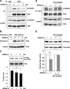
RGMa stimulation decreases the level of GTP-bound Ras through neogenin. A, B, Time course of Ras activity after RGMa-Fc (A) or Fc treatment (B) in N1E-115 cells. The N1E-115 cells were treated with RGMa-Fc or Fc for the indicated durations. Then, the cell lysates were incubated with GST-RBD, and bound GTP-Ras was detected using the anti-Ras antibody. The total Ras and neogenin expressions were detected by immunoblotting the cell lysates. C, Relative Ras activities after RGM-Fc (solid line) or Fc (dashed line) treatment in the N1E-115 cells. The relative Ras activity was determined on the basis of the amount of Ras bound to GST-RBD, which was normalized to the total amount of Ras in the cell lysates. The results are represented as means ± SD of three independent experiments. D, N1E-115 cells were transfected with neogenin shRNA or mutated neogenin shRNA, and the transfected cells were treated with RGMa-Fc for 5 min. The level of Ras activity was determined using the procedure described above. The relative Ras activities are expressed as the means ± SD of three independent experiments (graph). *p < 0.05 by Student's t test.
Figure 2.
Figure 2.
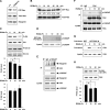
The involvement of p120GAP in the RGMa-induced inactivation of Ras, Akt, and Erk. A, RGMa-Fc treatment increases the interaction between p120GAP and GTP-Ras. The N1E-115 cells were treated with RGMa-Fc for the indicated durations. The cell lysates were incubated with GST-RBD, and the coprecipitated p120GAP and GTP-Ras were detected using anti-p120GAP and anti-Ras antibodies. The total p120GAP and Ras were detected by immunoblotting the cell lysates. B, C, The expression of the dominant-negative p120GAP (GAP-N) and the RNA interference (RNAi) knockdown of p120GAP inhibited RGMa-induced Ras inactivation. The N1E-115 cells were transfected with GAP-N-Myc or a mock plasmid (B) or p120GAP shRNA or mutated p120GAP shRNA (C), and they were treated with RGMa-Fc for 5 min. The levels of Ras activity were determined as described in Figure 1. The relative Ras activities are represented as means ± SD of three independent experiments (graph). *p < 0.05 by Student's t test. D, RGMa-Fc treatment decreased the phosphorylation of Akt and Erk in the N1E-115 cells. The N1E-115 cells were treated with RGMa-Fc for the indicated durations. The phosphorylation levels of Akt and Erk in the total cell lysates were analyzed by using anti-P-Akt and anti-P-Erk antibodies. The total Akt and Erk expressions were detected by anti-Akt and anti-Erk antibodies. The graphs indicate the relative P-Akt and P-Erk levels that were determined by the amount of P-Akt or P-Erk normalized to the total amounts of Akt or Erk, respectively, in the cell lysates. The results are represented as means ± SD of three independent experiments. *p < 0.05, **p < 0.01 by Student's t test. E, RNAi knockdown of p120GAP inhibited the RGMa-induced dephosphorylation of Akt and Erk. The N1E-115 cells were transfected with p120GAP shRNA or mutated p120GAP shRNA, and they were treated with RGMa-Fc for 5 min and lysed. The levels of P-Akt and P-Erk were determined as described in D. The relative P-Akt and P-Erk levels are represented as means ± SD of three independent experiments (graph). *p < 0.05, **p < 0.01 by Student's t test.
Figure 3.
Figure 3.
RGMa stimulation induces the dissociation of p120GAP from FAK through the dephosphorylation of FAK at Tyr-397. A, RGMa-Fc treatment decreased the interaction between FAK and p120GAP. The N1E-115 cells were treated with RGMa-Fc for the indicated durations and lysed. Then, the cell lysates were immunoprecipitated with anti-FAK antibody or control IgG, and the coprecipitated p120GAP was detected using the anti-p120GAP antibody. B, RGMa-Fc treatment decreased the interaction between FAK and GST-GAP-N. The N1E-115 cells were treated with RGMa-Fc for the indicated durations. Then, the cell lysates were incubated with GST-GAP-N, and the coprecipitated FAK was detected using an anti-FAK antibody. C, RGMa-Fc treatment decreased the Tyr-397 phosphorylation of FAK. The N1E-115 cells were treated with RGMa-Fc for the indicated durations. The Tyr-397 phosphorylation level of FAK in the total cell lysates was analyzed by using the anti-P-FAK (Tyr-397) antibody. The graph indicates the relative P-FAK level determined by the amount of P-FAK normalized to the total amount of FAK in the cell lysates. The results are expressed as means ± SD of three independent experiments (graph). *p < 0.05 by Student's t test. D, E, treatment with tyrosine phosphatase inhibitor prevented the RGMa-induced dissociation of p120GAP from FAK and the Ras inactivation. The N1E-115 cells were pretreated with 30 μm pervanadate for 10 min, followed by treatment with RGMa-Fc for 5 min. The cell lysates were immunoprecipitated with anti-FAK antibody, and the coprecipitated p120GAP was detected using the anti-p120GAP antibody. Ras activity was also analyzed by the GST-RBD pull-down assay, as described in Figure 1. The relative Ras activities are represented as the means ± SD of three independent experiments (graph). *p < 0.05 by Student's t test.
Figure 4.
Figure 4.
FAK mediates the interaction between p120GAP and neogenin. A, RGMa-Fc treatment did not influence the interaction between FAK and neogenin. The N1E-115 cells were treated with RGMa-Fc for the indicated durations and lysed. Then, the cell lysates were immunoprecipitated with the anti-neogenin antibody, and the coprecipitated FAK was detected using the anti-FAK antibody. B, SH2 domain of p120GAP interacts with neogenin and FAK. Cell lysates from the neogenin-VSV-expressing HEK293T cells were incubated with GST, GST-GAP-N, GST-SH2, or GST-SH3 bound to glutathione-Sepharose. The coprecipitated neogenin and FAK were analyzed by immunoblotting with the anti-VSV and anti-FAK antibodies. GST-fusion proteins were detected by Coomassie brilliant blue (CBB) staining. C, Expression of FAK WT, but not of the Y397F mutant, increased the amounts of neogenin and FAK precipitated with GST-GAP-N. The HEK293T cells were transfected with the indicated plasmids and lysed. The cell lysates were incubated with GST-GAP-N bound to glutathione-Sepharose. The coprecipitated neogenin and FAK were analyzed by immunoblotting with anti-VSV, anti-FAK, and anti-HA antibodies. Endo. FAK, Endogenous FAK. D, SFK inhibition did not influence the amounts of neogenin and FAK precipitated with GST-GAP-N. The HEK293T cells expressing neogenin-VSV and HA-FAK were treated with 5 μm PP2 or 5 μm PP3 for 30 min and lysed. The cell lysates were incubated with GST-GAP-N bound to glutathione-Sepharose. The coprecipitated neogenin and FAK were analyzed by immunoblotting with anti-VSV and anti-FAK antibodies. The phosphorylation levels of FAK at Tyr-397 and Tyr-861 in the total cell lysates were analyzed by anti-P-FAK (Tyr-397) and anti-P-FAK (Tyr-861) antibodies. E, FAK directly interacted with GST-GAP-N. Recombinant active FAK was incubated with GST or GST-GAP-N bound to glutathione-Sepharose. The precipitated FAK, GST, and GST-GAP-N proteins were detected by CBB staining. F, Neogenin, FAK, and GST-GAP-N form the triple complex. Cell lysates from the HEK293T cells transfected with neogenin-VSV or a mock plasmid were separated by SDS-PAGE, and subjected to Far Western blotting by using GST-GAP-N together with or without recombinant active FAK. Only the GST-GAP-N mixed with active FAK bound to neogenin-VSV. G, RGMa-Fc does not influence the interaction between p120GAP and neogenin. The N1E-115 cells were treated with RGMa-Fc for 5 min and lysed. Then, the cell lysates were immunoprecipitated with the anti-p120GAP antibody, and the coprecipitated neogenin was detected using the anti-neogenin antibody.
Figure 5.
Figure 5.
The involvement of p120GAP-mediated Ras inactivation in RGMa signaling in cortical neurons. A, RGMa treatment induced inactivation of Ras. Cortical neurons were treated with RGMa-Fc or Fc for 30 min. The levels of Ras activity were determined by using the procedure described in Figure 1. B, The time course of Ras activity after RGMa-Fc treatment in cortical neurons. The relative Ras activities are shown below the top panel. C, RGMa-Fc treatment decreased the phosphorylation of Akt, but not of Erk. The cortical neurons were treated with RGMa-Fc for the indicated durations and lysed. The levels of P-Akt and P-Erk were determined as described in Figure 2D. The relative P-Akt and P-Erk levels are represented as means ± SD of three independent experiments (graph). *p < 0.05 by Student's t test. D, RGMa-Fc treatment increased the interaction between p120GAP and GTP-Ras. The cortical neurons were treated with RGMa-Fc for the indicated durations. The cell lysates were incubated with GST-RBD, and the p120GAP coprecipitated with GTP-Ras was detected by using the anti-p120GAP antibody. The total p120GAP expression was detected by immunoblotting the cell lysates. E, Endogenous p120GAP interacts with neogenin and FAK. The lysates from cortical neurons were used for immunoprecipitation with the anti-p120GAP antibody or control IgG. The whole-cell lysates and the immunoprecipitates were probed with anti-neogenin, anti-FAK, and anti-p120GAP antibodies, as indicated. F, RGMa-Fc treatment decreased the interaction between FAK and p120GAP. The cortical neurons were treated with RGMa-Fc for the indicated durations and lysed. Then, the cell lysates were immunoprecipitated with anti-FAK antibody or control IgG, and the coprecipitated p120GAP was detected using the anti-p120GAP antibody. G, RGMa-Fc treatment decreased the interaction between FAK and GST-GAP-N. The cortical neurons were treated with RGMa-Fc for the indicated durations. Then, the cell lysates were incubated with GST-GAP-N, and the coprecipitated FAK was detected using the anti-FAK antibody. H, RGMa-Fc treatment decreased the phosphorylation of FAK at Tyr-397. The cortical neurons were treated with RGMa-Fc for the indicated durations. The phosphorylation level of FAK at Tyr-397 in the total cell lysates was analyzed by using the anti-P-FAK (Tyr-397) antibody. The graph indicates the relative P-FAK levels that were determined as described in Figure 3C. The results are expressed as means ± SD of three independent experiments (graph). *p < 0.05 by Student's t test.
Figure 6.
Figure 6.
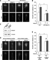
Ras inactivation mediated by p120GAP is required for RGMa-dependent growth cone collapse. A, The expression of constitutively active Ras prevented RGMa-induced growth cone collapse. The cortical neurons were cotransfected with RasV12 or a mock plasmid along with GFP. At 60 h after the transfection, the neurons were treated with RGMa-Fc or Fc for 30 min, fixed, and stained with phalloidin for visualizing F-actin. B, The percentages of the collapsed growth cones. The values are represented as the mean ± SD of three independent experiments (at least 70 growth cones in each experiment). *p < 0.01 by two-way ANOVA with Bonferroni's post hoc test. C, The effect of p120GAP shRNA on the expression of p120GAP in cortical neurons. The cortical neurons transfected with p120GAP shRNA or mutated p120GAP shRNA were cultured for 60 h and lysed. The expressions of p120GAP and α-tubulin were detected with anti-p120GAP and anti-α-tubulin antibodies, respectively. D, p120GAP knockdown prevented RGMa-induced growth cone collapse. The cortical neurons were cotransfected with p120GAP shRNA or mutated p120GAP shRNA along with GFP. At 60 h after the transfection, the neurons were treated with RGMa-Fc or Fc for 30 min, fixed, and stained with phalloidin for F-actin. E, The percentages of the collapsed growth cones. The values are represented as the means ± SD of three independent experiments (at least 70 growth cones in each experiment). *p < 0.01 by two-way ANOVA with Bonferroni's post hoc test.
Figure 7.
Figure 7.
p120GAP is required for RGMa-induced neurite outgrowth inhibition. A, p120GAP knockdown prevented RGMa-Fc-induced neurite outgrowth inhibition. The cortical neurons were transfected with p120GAP siRNA or control siRNA. At 24 h after the transfection, the neurons were replated and cultured with RGMa-Fc or Fc for 24 h. Then, the neurons were fixed and immunostained with the anti-βIII-tubulin antibody. Scale bar, 100 μm. B, Mean length of the longest neurite per neuron. The values are represented as the means ± SEM from at least 100 neurons for each population. *p < 0.01 by Student's t test. C, p120GAP knockdown prevented the neurite outgrowth inhibition induced by membrane-bound RGMa. The cortical neurons were transfected with p120GAP siRNA or control siRNA and cultured for 36 h on CHO cells (control) or RGMa-CHO cells (RGMa). Then, the neurons were fixed and immunostained with the anti-βIII-tubulin antibody. Scale bar, 100 μm. D, Mean length of the longest neurite per neuron. The values are represented as the mean ± SEM for at least 100 neurons for each population. *p < 0.01 by Student's t test.
Figure 8.
Figure 8.
Akt, but not Erk, mediates the RGMa-induced growth cone collapse. A, The effects of the inhibition of PI3 kinase or mitogen-activated protein kinase kinase (MEK) on the phosphorylation levels of Akt or Erk in the cortical neurons. The cortical neurons were treated with 100 nm wortmannin (Wort), 10 μm U0126, or DMSO for 30 min and lysed. The phosphorylation levels of Akt and Erk in the total cell lysates were analyzed by using anti-P-Akt and P-Erk antibodies. The total expressions of Akt and Erk were detected by the anti-Akt and anti-Erk antibodies. B, PI3 kinase inhibition, but not MEK inhibition, induced growth cone collapse in cortical neurons. The cortical neurons were cultured for 48 h and treated with 100 nm wortmannin, 10 μm U0126, or DMSO for 30 min. Then, the neurons were fixed and stained with phalloidin for detecting F-actin. C, The percentages of the collapsed growth cones after treatment with wortmannin, U0126, or DMSO. D, Expression of constitutively active Akt (Akt-myr) increases the amount of P-Akt, but it does not influence the amount of P-Erk. The cortical neurons were transfected with Akt-myr or a mock plasmid and cultured for 60 h. The levels of P-Akt and P-Erk were analyzed as described in A. E, Expression of Akt-myr prevents RGMa-induced growth cone collapse. The cortical neurons were cotransfected with Akt-myr or a mock plasmid along with GFP. At 60 h after the transfection, the neurons were treated with RGMa-Fc or Fc for 30 min, fixed, and stained with phalloidin. F, The percentages of collapsed growth cones. The values are represented as the means ± SD of three independent experiments (at least 70 growth cones in each experiment). *p < 0.01 by two-way ANOVA with Bonferroni's post hoc test.
Figure 9.
Figure 9.
Treatment with tyrosine phosphatase inhibitor prevents RGMa-induced growth cone collapse and FAK dephosphorylation. A, Cortical neurons were cultured for 48 h and pretreated with 1 mm orthovanadate for 30 min, which was followed by treatment with RGMa-Fc or Fc for 30 min. Then, the neurons were fixed and stained with phalloidin. B, The percentages of the collapsed growth cones. The values are represented as the means ± SD of three independent experiments (at least 70 growth cones in each experiment). *p < 0.01 by two-way ANOVA with Bonferroni's post hoc test. C, Cortical neurons were cultured for 48 h and pretreated with 1 mm orthovanadate for 30 min, followed by treatment with RGMa-Fc for 10 min. The Tyr-397 phosphorylation level of FAK in the total cell lysates was analyzed by using the anti-P-FAK (Tyr-397) antibody. The graph indicates the relative P-FAK level that was determined by using the procedure described in Figure 3C. The results are represented as the means ± SD of three independent experiments (graph). *p < 0.05 by Student's t test.
Figure 10.
Figure 10.
A diagram of the proposed mechanism of action of p120GAP in RGMa signaling. RGMa binding to neogenin accelerates the dissociation of p120GAP from FAK by dephosphorylating FAK at Tyr-397 via unidentified protein tyrosine phosphatase(s). The interaction between GTP-Ras and the p120GAP released from FAK mediates the inactivation of Ras and its downstream effector Akt. The inactivation of Ras and Akt contributes to RGMa-mediated repulsive function, including growth cone collapse and neurite outgrowth inhibition.

Similar articles

Cited by

References

    1. Boguski MS, McCormick F. Proteins regulating Ras and its relatives. Nature. 1993;366:643–654. - PubMed
    1. Brinks H, Conrad S, Vogt J, Oldekamp J, Sierra A, Deitinghoff L, Bechmann I, Alvarez-Bolado G, Heimrich B, Monnier PP, Mueller BK, Skutella T. The repulsive guidance molecule RGMa is involved in the formation of afferent connections in the dentate gyrus. J Neurosci. 2004;24:3862–3869. - PMC - PubMed
    1. Brouns MR, Matheson SF, Settleman J. p190 RhoGAP is the principal Src substrate in brain and regulates axon outgrowth, guidance and fasciculation. Nat Cell Biol. 2001;3:361–367. - PubMed
    1. Calalb MB, Zhang X, Polte TR, Hanks SK. Focal adhesion kinase tyrosine-861 is a major site of phosphorylation by Src. Biochem Biophys Res Commun. 1996;228:662–668. - PubMed
    1. Conrad S, Genth H, Hofmann F, Just I, Skutella T. Neogenin-RGMa signaling at the growth cone is bone morphogenetic protein-independent and involves RhoA, ROCK, and PKC. J Biol Chem. 2007;7:784–792. - PubMed

Publication types

MeSH terms