. 2009 May 20;29(20):6677-90.
doi: 10.1523/JNEUROSCI.4361-08.2009.

Lewis(x) and alpha2,3-sialyl glycans and their receptors TAG-1, Contactin, and L1 mediate CD24-dependent neurite outgrowth

Affiliations

Lewis(x) and alpha2,3-sialyl glycans and their receptors TAG-1, Contactin, and L1 mediate CD24-dependent neurite outgrowth

Annika Lieberoth et al. J Neurosci. .

Abstract

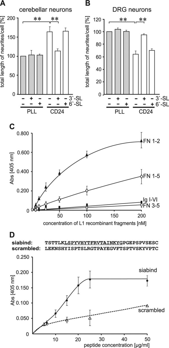
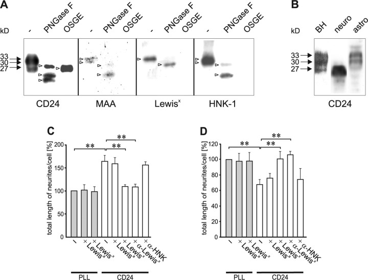
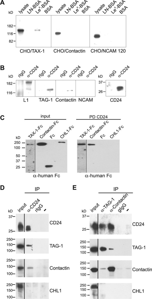
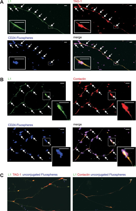
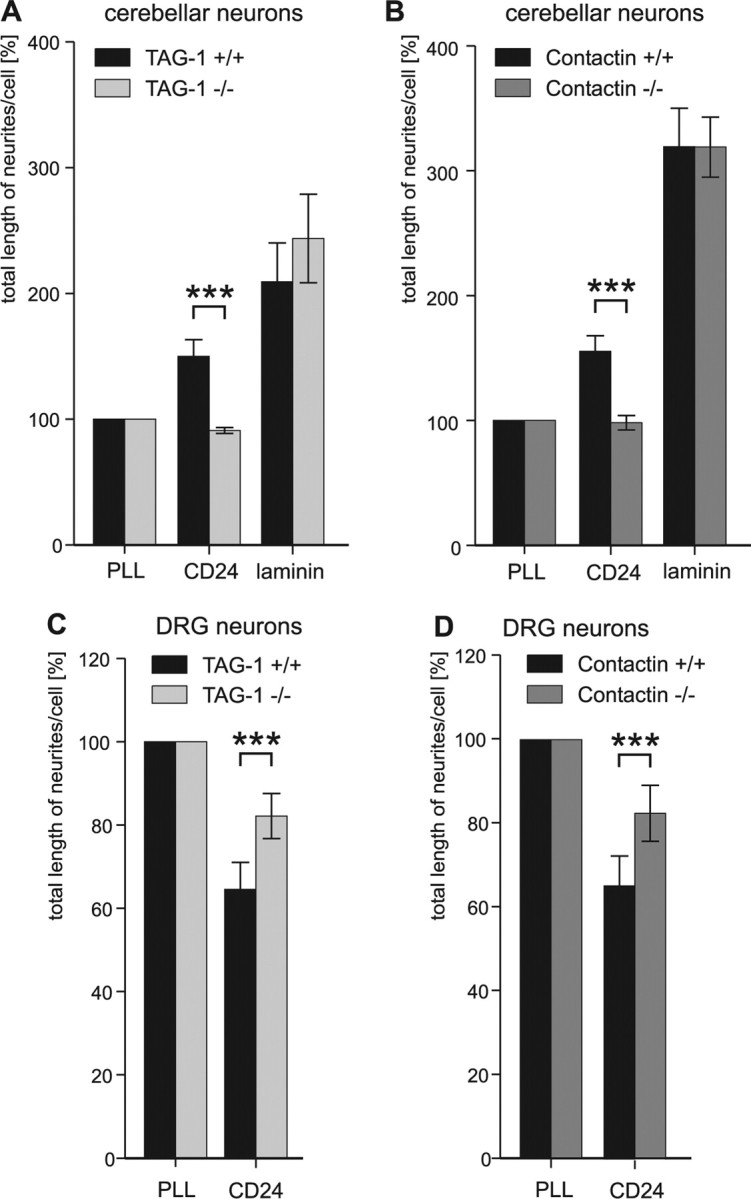
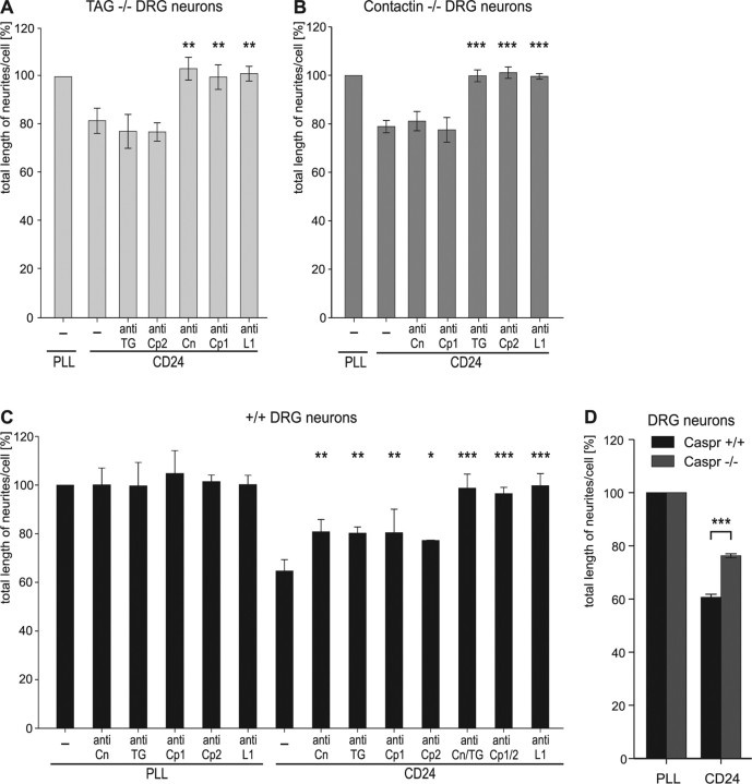
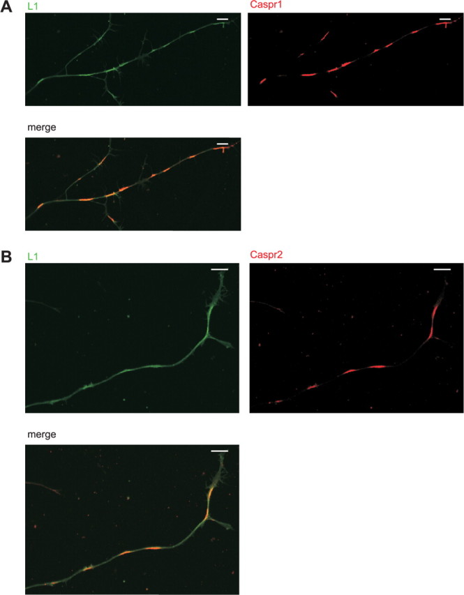
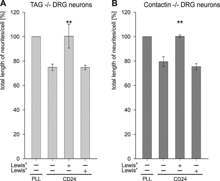
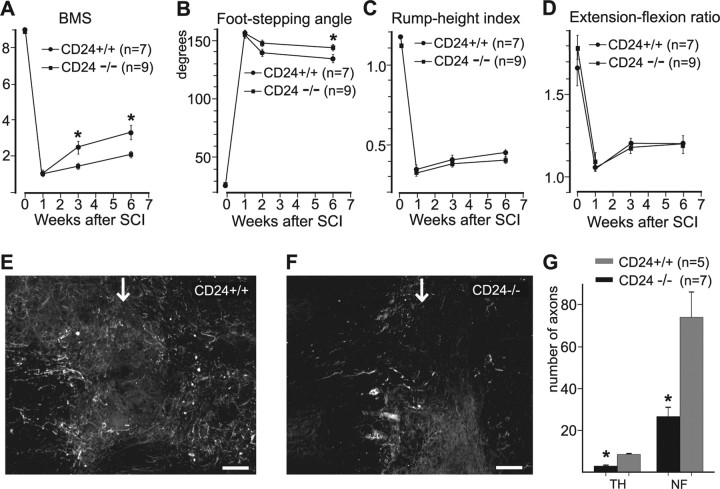
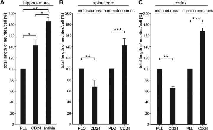
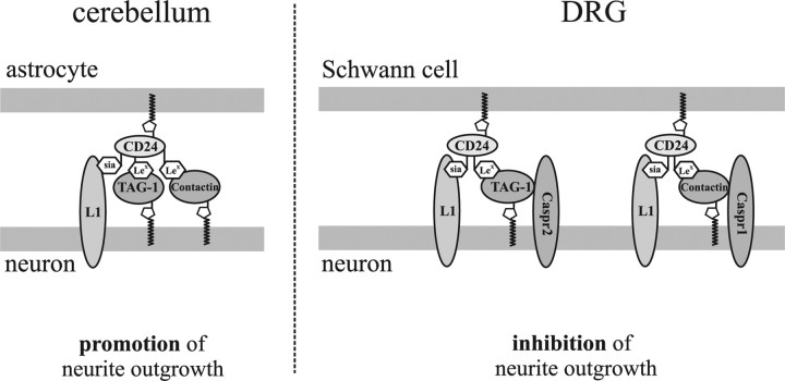
Although carbohydrates have been implicated in cell interactions in the nervous system, the molecular bases of their functions have remained largely obscure. Here, we show that promotion or inhibition of neurite outgrowth of cerebellar or dorsal root ganglion neurons, respectively, induced by the mucin-type adhesion molecule CD24 depends on alpha2,3-linked sialic acid and Lewis(x) present on glia-specific CD24 glycoforms. Alpha2,3-sialyl residues of CD24 bind to a structural motif in the first fibronectin type III domain of the adhesion molecule L1. Following the observation that the adhesion molecules TAG-1 and Contactin show sequence homologies with fucose-specific lectins, we obtained evidence that TAG-1 and Contactin mediate Lewis(x)-dependent CD24-induced effects on neurite outgrowth. Thus, L1, TAG-1, and Contactin function as lectin-like neuronal receptors. Their cis interactions with neighboring adhesion molecules, e.g., Caspr1 and Caspr2, and with their triggered signal transduction pathways elicit cell type-specific promotion or inhibition of neurite outgrowth induced by glial CD24 in a glycan-dependent trans interaction.


Figures

Figure 1.
Figure 1.
Sialic acid-dependent binding of L1 to CD24 affects neurite outgrowth. A, B, Cerebellar neurons (A) and DRG neurons (B) were grown on substrate-coated PLL or CD24 in the absence or presence of 3′-sialyl-N-acetyllactosamine (3′-SL) or 6′-sialyl-N-acetyllactosamine (6′-SL). Total length of neurites per cell was determined and shown as percentage of PLL control. Error bars indicate SD from three independent experiments. Bars marked by double asterisks (p < 0.01, Student's t test) are significantly different from the controls (PLL or CD24). C, D, Brain CD24 was substrate coated and incubated with different amounts of L1 fragments (C) and biotinylated L1-derived peptide (siabind) and its scrambled form (D). Amino acid sequences of peptides are shown (D) and underlined amino acids represent the putative sialic acid binding motif. Binding was evaluated by ELISA using L1 antibody (C) or streptavidin (D). Error bars indicate SD from at least three independent experiments. Abs, Absorbance.
Figure 2.
Figure 2.
CD24 glycosylation pattern and effects of Lewisx and HNK-1 glycans on CD24-induced neurite outgrowth. A, Mock-treated CD24 (−), and CD24 treated with PNGase F or OSGE was subjected to Western blot analysis using CD24, Lewisx, and HNK-1 antibodies or MAA. Arrows indicate the three CD24 glycoforms and arrowheads indicate the positions of individual bands after PNGase F, OSGE, or mock treatment. B, Mouse brain homogenate (BH), cell lysates of cultured cerebellar neurons (neuro), or astrocytes (astro) were analyzed using CD24 antibody. Arrows indicate the different CD24 glycoforms. Clear bands are not always visible, as separation of highly glycosylated glycoproteins is notoriously difficult. C, D, Cerebellar neurons (C) and DRG neurons (D) were grown on PLL or CD24 in absence (−) or presence of Lewisa, Lewisx, Lewisx antibody, or HNK-1 antibody. Total length of neurites per cell was determined and shown as percentage of PLL control. Error bars indicate SD from three independent experiments. Bars marked by double asterisks (p < 0.01, Student's t test) are significantly different from the control (PLL or CD24).
Figure 3.
Figure 3.
TAG-1 and Contactin bind to CD24 and are receptors for Lewisx. A, Lysates from CHO cells transfected with TAX-1 (CHO/TAX-1), Contactin (CHO/Contactin), or NCAM120 (CHO/NCAM120) were incubated with epoxy beads coated with Lewisx–BSA (Lex-BSA), N-acetyllactosamine–BSA (LN-BSA), and BSA. After pull-down, beads were analyzed by Western blotting using antibodies to TAG-1 (lanes 1–4), Contactin (lanes 5–8), or NCAM (lanes 9–12). B, Immunoprecipitates from brain homogenate using CD24 antibody (α-CD24) or control rat IgG (rIgG) were analyzed by Western blotting using antibodies against L1, TAG-1, Contactin, NCAM, or CD24. C, CD24-coupled epoxy beads were incubated with TAX-1-Fc, Contactin-Fc, CHL1-Fc, or Fc alone. After pull-down, beads (PD CD24) were analyzed by Western blotting using HRP-coupled anti-human antibody. D, E, Cerebellar tissue (input) was used for immunoprecipitations with CD24 with antibody (α-CD24), with control rat IgG (rIgG) or without antibody (−) or for immunoprecipitations with TAG-1 antibody (α-TAG-1), Contactin antibody (α-Contactin), or control goat IgG (gIgG) or without antibody (−). Immunoprecipitates were analyzed by Western blotting using antibodies against CD24, TAG-1, Contactin, or CHL1.
Figure 4.
Figure 4.
CD24 binds in trans to L1/TAG-1 or L1/Contactin clusters on cultured DRG neurons. A–C, Primary cultures of wild-type DRG neurons were incubated with CD24 coated (A, B) or unconjugated (C) Fluospheres and afterward immunofluorescent staining of L1 and TAG-1 (A, C) or L1 and Contactin (B, C) was performed. Arrows indicate clusters stained for CD24-Fluospheres, L1 and TAG-1, or Contactin. Areas in rectangles are magnified. Scale bars, 10 μm.
Figure 5.
Figure 5.
Involvement of TAG-1 and Contactin in CD24-induced neurite outgrowth. A–D, Cerebellar (A, B) and DRG (C, D) neurons from TAG-1-deficient (−/−) (A, C) or Contactin-deficient (−/−) (B, D) and wild-type littermate (+/+) mice were grown on PLL, CD24, or laminin. Total length of neurites per cell was determined and is shown as percentage of PLL control. Error bars indicate SD from three independent experiments. Bars marked by triple asterisks (p < 0.001) are significantly different from the wild-type control (Student's t test).
Figure 6.
Figure 6.
Synergistic effects of TAG-1/Caspr2 and Contactin/Caspr in CD24-mediated neurite outgrowth of DRG neurons. A–D, DRG neurons from TAG-1-deficient (−/−) (A), Contactin-deficient (−/−) (B), and wild-type littermate (+/+) (C) mice or DRG neurons from Caspr1-deficient (−/−) and wild-type littermate (+/+) mice (D) were grown on PLL or CD24 in the absence or presence of antibodies against TAG-1 (TG), Contactin (Cn), Caspr1 (Cp1), Caspr2 (Cp2), or L1 (A–C). Total length of neurites per cell was determined and is shown as percentage of PLL control. Error bars indicate SD from three independent experiments. Bars marked by single asterisks (p < 0.05), double asterisks (p < 0.01), and triple asterisks (p < 0.001) are significantly different from the wild-type control (Student's t test). A–C, Values for PLL were not affected by any antibody, as exemplified in C.
Figure 7.
Figure 7.
Caspr1 and Caspr2 colocalize with L1 on cultured DRG neurons. A, B, DRG neurons grown on PLL were labeled by immunofluorescence using antibodies to L1 and Caspr1 (A) or L1 and Caspr2 (B). Scale bars, 10 μm.
Figure 8.
Figure 8.
Lewisx-dependent effects of TAG-1 and Contactin in CD24-mediated neurite outgrowth of DRG neurons. A, B, DRG neurons from TAG-1-deficient (−/−) (A) and Contactin-deficient (−/−) (B) mice were grown on PLL or CD24 in the absence or presence of Lewisa or Lewisx. Total length of neurites per cell was determined and is shown as percentage of PLL control. Error bars indicate SD from three independent experiments. Bars marked by double asterisks (p < 0.01) are significantly different from the wild-type control (Student's t test). Values for PLL were not affected by any glycan, as exemplified in Figure 2D.
Figure 9.
Figure 9.
Regeneration after spinal cord injury in CD24+/+ and CD24−/− mice. A–D, Time course and degree of functional recovery in CD24−/− mice and CD24+/+ littermates after spinal cord injury. Shown are mean values ± SEM of open-field locomotion (BMS) scores (A), foot-stepping angles (B), rump-height indices (C), and extension-flexion ratios (D) before surgery (day 0) and at 1, 3, and 6 weeks after injury. Numbers of animals are given in parentheses; asterisks indicate significant differences between group mean values at a given time point (p < 0.05, one-way ANOVA for repeated measurements with Tukey post hoc test). E, F, Representative images of immunofluorescent stainings for neurofilament at the lesion epicenter (arrow) in CD24+/+ littermate (E) and CD24−/− (F) mice. Left is rostral, right is caudal. G, Mean numbers of TH-positive and NF-positive fibers crossing an arbitrary border 250 μm caudally from the lesion site at 6 weeks after the lesion (*p < 0.05; Wilcoxon–Mann–Whitney test).
Figure 10.
Figure 10.
CD24-mediated neurite outgrowth of hippocampal, cortical, and spinal cord neurons. A, Hippocampal neurons from wild-type mice were grown on PLL, CD24, or laminin. B, Wild-type spinal cord neurons were grown on PLO or CD24. The culture contains round non-motoneurons with a diameter of ∼10 μm and motoneurons having a large triangular cell body with an edge length of 15–20 μm. C, Wild-type cortical neurons were grown on PLL or CD24. The culture contains non-motoneurons and motoneurons. A–C, Total length of neurites per cell was determined and is shown as percentage of the PLL or PLO controls. Error bars indicate SD from three independent experiments. Bars marked by asterisks (p < 0.05) or double asterisks (p < 0.01) are significantly different from the control (PLL, PLO, or laminin; Student's t test).
Figure 11.
Figure 11.
Scheme of a working model: role of Lewisx and α2,3-linked sialic acid in modulating glia–neuron interactions. L1 on neurons binds to α2,3-linked sialic acid carried by glia-derived CD24, and neuronal TAG-1 and Contactin bind to Lewisx carried by glia-derived CD24. In the case of cerebellar neurons, a complex formed by neuronal L1, TAG-1, and Contactin leads to signal transduction promoting neurite outgrowth. Inhibition of neurite outgrowth in DRG neurons is induced by two independent complexes of either L1, TAG-1, and Caspr2 or L1, Contactin, and Caspr1.

Similar articles

Cited by

References

    1. Apostolova I, Irintchev A, Schachner M. Tenascin-R restricts posttraumatic remodeling of motoneuron innervation and functional recovery after spinal cord injury in adult mice. J Neurosci. 2006;26:7849–7859. - PMC - PubMed
    1. Appel F, Holm J, Conscience JF, Schachner M. Several extracellular domains of the neural cell adhesion molecule L1 are involved in neurite outgrowth and cell body adhesion. J Neurosci. 1993;13:4764–4775. - PMC - PubMed
    1. Basso DM, Fisher LC, Anderson AJ, Jakeman LB, McTigue DM, Popovich PG. Basso Mouse Scale for locomotion detects differences in recovery after spinal cord injury in five common mouse strains. J Neurotrauma. 2006;23:635–659. - PubMed
    1. Berglund EO, Murai KK, Fredette B, Sekerková G, Marturano B, Weber L, Mugnaini E, Ranscht B. Ataxia and abnormal cerebellar microorganization in mice with ablated Contactin gene expression. Neuron. 1999;24:739–750. - PubMed
    1. Bhat MA, Rios JC, Lu Y, Garcia-Fresco GP, Ching W, St Martin M, Li J, Einheber S, Chesler M, Rosenbluth J, Salzer JL, Bellen HJ. Axon-glia interactions and the domain organization of myelinated axons requires neurexin IV/Caspr/Paranodin. Neuron. 2001;30:369–383. - PubMed

Publication types

MeSH terms