

Solution structure of eggcase silk protein and its implications for silk fiber formation
- PMID: 19458259
- PMCID: PMC2690042
- DOI: 10.1073/pnas.0813255106
Solution structure of eggcase silk protein and its implications for silk fiber formation
Abstract
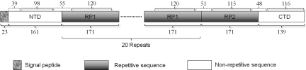
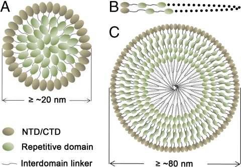
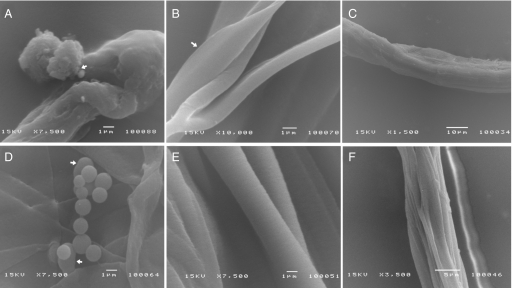
Spider silks are renowned for their excellent mechanical properties and biomimetic and industrial potentials. They are formed from the natural refolding of water-soluble fibroins with alpha-helical and random coil structures in silk glands into insoluble fibers with mainly beta-structures. The structures of the fibroins at atomic resolution and silk formation mechanism remain largely unknown. Here, we report the 3D structures of individual domains of a approximately 366-kDa eggcase silk protein that consists of 20 identical type 1 repetitive domains, one type 2 repetitive domain, and conserved nonrepetitive N- and C-terminal domains. The structures of the individual domains in solution were determined by using NMR techniques. The domain interactions were investigated by NMR and dynamic light-scattering techniques. The formation of micelles and macroscopic fibers from the domains was examined by electron microscopy. We find that either of the terminal domains covalently linked with at least one repetitive domain spontaneously forms micelle-like structures and can be further transformed into fibers at > or = 37 degrees C and a protein concentration of > 0.1 wt%. Our biophysical and biochemical experiments indicate that the less hydrophilic terminal domains initiate the assembly of the proteins and form the outer layer of the micelles whereas the more hydrophilic repetitive domains are embedded inside to ensure the formation of the micelle-like structures that are the essential intermediates in silk formation. Our results establish the roles of individual silk protein domains in fiber formation and provide the basis for designing miniature fibroins for producing artificial silks.
Conflict of interest statement
The authors declare no conflict of interest.
Figures


References
-
- Foelix RF. The Biology of Spiders. New York: Oxford Univ Press; 1996.
-
- Vollrath F, Knight DP. Liquid crystalline spinning of spider silk. Nature. 2001;410:541–548. - PubMed
-
- Guerette PA, Ginzinger DG, Weber BHF, Gosline JM. Silk properties determined by gland-specific expression of a spider fibroin gene family. Science. 1996;272:112–115. - PubMed
-
- Huang W, et al. Characterization and expression of a cDNA encoding a tubuliform silk protein of the golden web spider Nephila antipodiana. Biochimie. 2006;88:849–858. - PubMed
Publication types
MeSH terms
Substances
Associated data
- Actions
- Actions
- Actions
- Actions
- Actions
LinkOut - more resources
Full Text Sources
Other Literature Sources
Molecular Biology Databases
