. 2009 Jul;297(1):R210-7.
doi: 10.1152/ajpregu.90989.2008. Epub 2009 May 20.

The posterior vermis of the cerebellum selectively inhibits 10-Hz sympathetic nerve discharge in anesthetized cats

Affiliations

The posterior vermis of the cerebellum selectively inhibits 10-Hz sympathetic nerve discharge in anesthetized cats

Susan M Barman et al. Am J Physiol Regul Integr Comp Physiol. 2009 Jul.

Abstract

We studied the changes in inferior cardiac sympathetic nerve discharge (SND) and mean arterial pressure (MAP) produced by aspiration or chemical inactivation (muscimol microinjection) of lobule IX (uvula) of the posterior vermis of the cerebellum in baroreceptor-denervated and baroreceptor-innervated cats anesthetized with urethane. Autospectral analysis was used to decompose SND into its frequency components. Special attention was paid to the question of whether the experimental procedures affected the rhythmic (10-Hz and cardiac-related) components of SND. Aspiration or chemical inactivation of lobule IX produced an approximately three-fold increase in the 10-Hz rhythmic component of SND (P < or = 0.05) in baroreceptor-denervated cats. Total power (0- to 20-Hz band) was unchanged. Despite the absence of a change in total power in SND, there was a statistically significant increase in MAP. In baroreceptor-innervated cats, neither aspiration nor chemical inactivation of the uvula caused a significant change in cardiac-related or total power in SND or MAP. These results are the first to demonstrate a role of cerebellar cortical neurons of the posterior vermis in regulating the frequency composition of naturally occurring SND. Specifically, these neurons selectively inhibit the 10-Hz rhythm-generating network in baroreceptor-denervated, urethane-anesthetized cats. The functional implications of these findings are discussed.


Figures

Fig. 1.
Fig. 1.
Effects of aspiration of the posterior vermis of the cerebellum on arterial pressure (AP) and left inferior cardiac sympathetic nerve discharge (SND) in a baroreceptor-denervated cat. A: oscillographic records (top to bottom) show AP (mmHg), SND, end tidal CO2 (%), and time base (10 s/division). Aspiration was performed over a 12-s period (see bars above time base). Horizontal bars (1 and 2) above SND are presented on an expanded time base (B). Vertical calibration, 100 μV. C: autospectra of SND before (black trace) and 3–5 min (gray trace) after ablation of the posterior vermis. Autospectra in this and subsequent figures are based on 47 5-s windows with 50% overlap and have a frequency resolution of 0.2 Hz per bin.
Fig. 2.
Fig. 2.
Midsagittal sections of the cerebellum showing extent of ablation of the posterior vermis in three cats. Drawings are based on those of Larsell (16, 17) and Ito (15). A: drawing of a midsagittal section of a normal (no ablation) cerebellum showing lobules I–X and sublobules IX, a–d in the uvula. B: black region marks the portion of the cerebellum (sublobules IX, b–d) removed in one cat (same as in Fig. 1). C: removal of superficial portions of sublobules IX, a–c in another cat (same as in Fig. 3B). D: drawing showing the largest ablation performed in this series of experiments. In this case, ablation included all of lobule X and portions of sublobules IX, b–d. Calibration, 1 mm.
Fig. 3.
Fig. 3.
Removal of the posterior vermis enhanced 10-Hz activity in the inferior cardiac nerve in a baroreceptor-denervated cat. Autospectra of SND before (black traces) and after (gray traces) ablation of the posterior vermis.
Fig. 4.
Fig. 4.
Summary of the changes in SND and mean arterial pressure (MAP) produced by ablation or chemical inactivation (microinjection of muscimol ) of the posterior vermis. A: 10-Hz, 0- to 6-Hz, and total (0- to 20-Hz) power in SND before (Cont) and after aspiration of the posterior vermis (Cbx) in baroreceptor-denervated cats. B: same, for effects of microinjection of muscimol (Musc) into the posterior vermis in baroreceptor-denervated cats. C: cardiac-related, 0- to 6-Hz, and total (0- to 20-Hz) power in SND before (Cont) and after aspiration of the posterior vermis (Cbx) in baroreceptor-innervated cats. D: same, for effects of microinjection of muscimol (Musc) into the posterior vermis in the same baroreceptor-innervated cats. The insets show MAP before and after the manipulations. Values are means ± SE. *Statistically different from before aspiration or chemical inactivation (P ≤ 0.05; paired t-test). The numbers in the bars refer to the number of experiments.
Fig. 5.
Fig. 5.
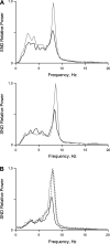
Comparison of effects of chemical inactivation (muscimol microinjection) and aspiration of the posterior vermis in the same baroreceptor-denervated cats. A, top: autospectra of SND before (black trace) and after (gray trace) microinjection of muscimol into the posterior vermis. A, bottom: autospectrum of SND 2 h later (black trace) at which time the power and frequency composition of SND had returned to near control and after subsequent aspiration of the posterior vermis (gray trace). B: autospectra of SND before (solid black trace) and after (gray trace) microinjection of muscimol into the posterior vermis and after ablation of the posterior vermis 15 min later (dotted black trace).
Fig. 6.
Fig. 6.
Effects of chemical inactivation (muscimol microinjection) and aspiration of the posterior vermis in a baroreceptor-innervated cat. A: autospectra of SND before (black trace) and after (gray trace) microinjection of muscimol into the posterior vermis. B: autospectra of SND before (black traces) and after (gray trace) aspiration of the posterior vermis 30 min later.

References

    1. Barman SM, Gebber GL. Tonic sympathoinhibition in the baroreceptor-denervated cat. Proc Soc Exp Biol Med 157: 646–653, 1978. - PubMed
    1. Barman SM, Gebber GL. 'Rapid' rhythmic discharges of sympathetic nerves: Sources, mechanisms of generation, and physiological relevance. J Biol Rhythms 15: 365–379, 2000. - PubMed
    1. Barman SM, Gebber GL, Kitchens H. Rostral dorsolateral pontine neurons with sympathetic nerve-related activity. Am J Physiol Heart Circ Physiol 276: H401–H412, 1999. - PubMed
    1. Barman SM, Gebber GL, Zhong S. The 10-Hz rhythm in sympathetic nerve discharge. Am J Physiol Regul Integr Comp Physiol 262: R1006–R1014, 1992. - PubMed
    1. Barman SM, Kitchens HL, Leckow AB, Gebber GL. Pontine neurons are elements of the network responsible for the 10-Hz rhythm in sympathetic nerve discharge. Am J Physiol Heart Circ Physiol 273: H1909–H1919, 1997. - PubMed

Publication types

MeSH terms

LinkOut - more resources