. 2009 Sep;81(3):571-9.
doi: 10.1095/biolreprod.109.075952. Epub 2009 May 20.

Activation and repression domains within the promoter of the rat cathepsin L gene orchestrate sertoli cell-specific and stage-specific gene transcription in transgenic mice

Affiliations

Activation and repression domains within the promoter of the rat cathepsin L gene orchestrate sertoli cell-specific and stage-specific gene transcription in transgenic mice

Thomas Visone et al. Biol Reprod. 2009 Sep.

Abstract

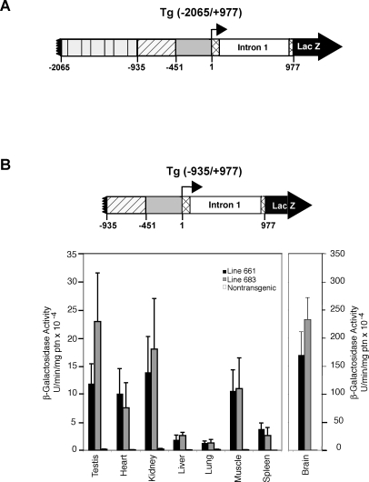
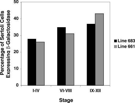
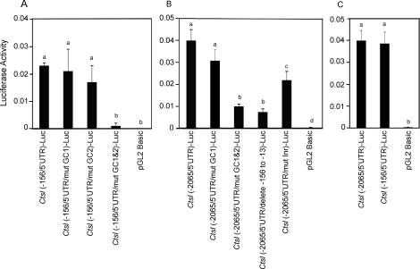
In murine testes, only Sertoli cells express the cathepsin L (Ctsl) gene, and this expression is restricted to stages V-VIII of the cycle. Our previous transgenic analysis of Tg (-2065/+977) demonstrated that this expression is regulated by a approximately 2-kb promoter. To begin to elucidate this regulation, we analyzed the in vivo expression of two new transgenes, Tg (-935/+977) and Tg (-451/+977). Tg (-935/+977) was expressed by Sertoli cells but, in contrast to Tg (-2065/+977), was expressed at all stages of the cycle, by spermatocytes, by the vascular endothelium, and by seven other organs. Tg (-451/+977) was not expressed by Sertoli cells but by spermatogenic cells and by the brain. Lack of expression of Tg (-451/+977) by Sertoli cells was not due to a lack of essential cis-acting elements. Transient transfection analysis of primary cultures of mature rat Sertoli cells demonstrated that in mature Sertoli cells, most of the activity of the Ctsl promoter is accounted for by one of two redundant upstream GC motifs and an Initiator that are within 100 bp of the transcription start site. We conclude that transcriptional repressors upstream from nucleotide -935 of the rat Ctsl gene restrict testicular expression of this gene to Sertoli cells at stages V-VIII. At these stages, transcriptional activators located between nucleotides -935 and -452 promote access of the transcriptional machinery to the two GC boxes and to the Initiator. Thus, upstream repressors and activators as well as cis-acting elements near the transcription start site control stage-specific Ctsl transcription by Sertoli cells.


Figures

FIG. 1.
FIG. 1.
Expression of Tg (−935/+977) in various organs of transgenic mice and schematic representations of Tg (−2065/+977) and Tg (−935/+977). A) Tg (−2065/+977) has been shown previously to drive both stage-specific and Sertoli cell-specific gene expression in transgenic mice [12]. The promoter contains 2065 nucleotides upstream of the TSS (black box), the first exon (74 nucleotides; hatched box), the first intron (892 nucleotides; white box), and the first 11 nucleotides of exon 2 (hatched box) up to the ATG initiation codon at nucleotide +978. The position of the TSS is indicated by the bent arrow. The numbering is relative to the Ctsl TSS, which is designated +1. For comparison with the results presented in this paper, the previously published data for expression of Tg (−2065/+977) in various organs are provided in Supplemental Table S2. B) β-Galactosidase enzymatic activity in testis and other organs of two lines of mice expressing the transgene Tg (−935/+977). Data (n = 3; means ± SEM) are shown for transgenic mice in line 661 and line 683, as well as for control, nontransgenic mice. In both transgenic lines, β-galactosidase activity in each organ was statistically greater than the activity in the same organ of control mice. The mice used for this analysis were 90 to 150 days old. ptn, protein.
FIG. 2.
FIG. 2.
Expression of Tg (−935/+977) in specific testicular cell types. To identify the cells expressing Tg (−935/+977), fixed testes were incubated in X-gal, which is metabolized by β-galactosidase to produce an insoluble blue reaction product. The testes were then embedded in paraffin, and 8-μm sections were cut and counterstained with nuclear fast red. A) Expression of the transgene at stage I or II. Arrow points to a Sertoli cell nucleus. B) Expression of the transgene at stage VII. Arrow points to a Sertoli cell nucleus; arrowhead points to a preleptotene spermatocyte nucleus. C) Expression of the transgene at stage X. Arrowhead points to a spermatocyte nucleus. D) Expression of the transgene at stage XI. Arrow points to a Sertoli cell nucleus. E) Lack of β-galactosidase activity in a stage IV or V seminiferous tubule of a control, nontransgenic mouse. Arrow points to a Sertoli cell nucleus. F) Expression of Tg (−935/+977) in a testicular blood vessel. Bars = 10 μm on the original tissue section.
FIG. 3.
FIG. 3.
The transgene Tg (−935/+977) is expressed by Sertoli cells at all stages of the cycle of the seminiferous epithelium. One testis from line 661 and one testis from line 683 were incubated in X-gal, embedded in paraffin, and serially sectioned. β-Galactosidase-positive Sertoli cells in stage I–V, VI–VIII, or IX–XII tubules were counted. These groups of stages occur with similar frequency within a mouse testis [21]. The numbers of β-galactosidase-expressing Sertoli cells at stages I–V, VI–VIII, or IX–XII were expressed as the percentage of all β-galactosidase-positive Sertoli cells in each testis. The mice used for this analysis were 90–100 days old.
FIG. 4.
FIG. 4.
β-Galactosidase enzymatic activity in testis and various other organs of two lines of mice expressing the transgene Tg (−451/+977). Data (n = 3; means ± SEM) are shown for transgenic mice in lines 251 and 263 as well as for control, nontransgenic mice. In both transgenic lines, β-galactosidase activity was statistically greater in testis and brain than in the comparable organs of control mice. ptn, protein.
FIG. 5.
FIG. 5.
Immunocytochemical localization of β-galactosidase in a testis of a Tg (−451/+977) mouse. A) Immunocytochemical localization of β-galactosidase in a testis section of a transgenic mouse. B) Lack of β-galactosidase antigen in a testis of a control, nontransgenic mouse. C) Higher magnification of cells expressing β-galactosidase in a transgenic testis. Intensely staining cells are pachytene spermatocytes. The binding of antibody to its antigen in the tissue sections was detected by development of a peroxidase reaction product. Images were taken using phase-contrast optics. Bars = 10 μm on the original tissue section.
FIG. 6.
FIG. 6.
β-Galactosidase activity in testes, interstitium, seminiferous tubules, germ cells, and Sertoli cells of mice carrying the Tg (−451/+977) or nontransgenic, control mice. Seminiferous tubules, spermatogenic cells, and interstitium were isolated and analyzed in a separate experiment from Sertoli cells. Tubules and cells were isolated from lines 251 and 263, and data (mean ± SEM) were pooled. A) Measurement of β-galactosidase activity in testes, interstitium, seminiferous tubules, and germ cells of transgenic mice. β-Galactosidase activity in germ cells isolated from control, wild-type mice was also measured. B) Measurement of β-galactosidase activity in testes of transgenic mice, in Sertoli cells of transgenic mice, and in Sertoli cells isolated from testes of control, nontransgenic mice. Data were obtained from cells isolated from two mice of line 263 and from cells isolated from one mouse of line 251. Data from both lines were similar, and data were pooled for purposes of statistical analysis. Means labeled by different letters differ statistically from the other means in that part of the experiment.
FIG. 7.
FIG. 7.
Identification of cis-acting regulatory elements that confer Ctsl promoter activity when transfected into Sertoli cells isolated from sexually mature rats. A) Demonstration that GC1 and GC2 have redundant activities when tested in the context of 156 bp of Ctsl promoter immediately upstream from the TSS. Mature rat Sertoli cells were transfected with one of the following reporter constructs: Ctsl (−156/5′ UTR)-Luc, Ctsl (−156/5′ UTR/mut GC1)-Luc, Ctsl (−156/5′ UTR/mut GC2)-Luc, Ctsl (−156/5′ UTR/mut GCs 1 and 2)-Luc, or the negative control plasmid, pGL2 Basic. B) Demonstration that GC1 and GC2 have redundant activities when tested in the context of 2065 bp of Ctsl promoter immediately upstream from the TSS and the demonstration that the Initiator (Inr) is required for full promoter activity. Mature Sertoli cells were transfected with one of the following Ctsl reporter constructs: Ctsl (−2065/5′ UTR)-Luc, Ctsl (−2065/5′ UTR/mut GC1)-Luc, Ctsl (−2065/5′ UTR/mut GCs 1 and 2)-Luc, Ctsl (−2065/5′ UTR/delete −156 to −13)-Luc, Ctsl (−2065/5′ UTR/mut Inr)-Luc, or pGL2 basic. C) The 156 bp of Ctsl promoter immediately upstream from the TSS exhibits maximal activity. Mature Sertoli cells were transfected with Ctsl (−2065/5′ UTR)-Luc, Ctsl (−156/5′ UTR)-Luc, or pGL2 basic. In all three experiments, potential differences in transfection efficiency were corrected by cotransfecting the Sertoli cells with pRL-CMV, which encodes Renilla luciferase. At 24 h after transfection, Sertoli cells were collected, luciferase activities were measured, and Luc activity was defined as firefly luciferase activity/Renilla luciferase activity. Data (mean ± SEM) were obtained from four independent replicates of each experiment. Means labeled with different letters differ statistically. GC1&2, GCs 1 and 2.
FIG. 8.
FIG. 8.
A proposed model to explain how stage-specific expression of the Ctsl gene in rat Sertoli cells is regulated. Research described in this paper and preceding papers [12, 16, 17] identifies four functional domains within the region that spans −2065 to +977 of the Ctsl gene. Domain II contains two redundant GC boxes, GC1 and GC2. We propose that access of SP transcription family members to these sites is regulated by cis-acting elements within domains III and IV. This model predicts that at stages VI–VIII, domain III recruits transcription factors that alone or in conjunction with coactivators relax the chromatin structure of domain II. We propose that this relaxation allows access of SP family members to GC1 and GC2. Expression of the Ctsl gene at stages VI–VIII is then enhanced by the action of domain I, the first intron. This model also predicts that at stages I–IV and IX–XIV, cis-acting elements in domain IV block the ability of domain III to relax the chromatin structure of domain II, rendering the GC boxes inaccessible to SP family members. Thus, we propose that stage-specific expression of the Ctsl gene by Sertoli cells results from the activities of upstream elements in domains III and IV that regulate the chromatin structure of domain II. GC1&2, GCs 1 and 2.

Similar articles

Cited by

References

    1. Behrens A, Genoud N, Naumann H, Rulicke T, Janett F, Heppner FL, Ledermann B, Aguzzi A.Absence of the prion protein homologue Doppel causes male sterility. EMBO J 2002; 21: 3652–3658.. - PMC - PubMed
    1. Brehm R, Zeiler M, Ruttinger C, Herde K, Kibschull M, Winterhager E, Willecke K, Guillou F, Lecureuil C, Steger K, Konrad L, Biermann K, et al. A sertoli cell-specific knockout of connexin43 prevents initiation of spermatogenesis. Am J Pathol 2007; 171: 19–31.. - PMC - PubMed
    1. Chang C, Chen YT, Yeh SD, Xu Q, Wang RS, Guillou F, Lardy H, Yeh S.Infertility with defective spermatogenesis and hypotestosteronemia in male mice lacking the androgen receptor in Sertoli cells. Proc Natl Acad Sci U S A 2004; 101: 6876–6881.. - PMC - PubMed
    1. Holdcraft RW, Braun RE.Androgen receptor function is required in Sertoli cells for the terminal differentiation of haploid spermatids. Development 2004; 131: 459–467.. - PubMed
    1. Wright WW, Smith L, Kerr C, Charron M.Mice that express enzymatically inactive cathepsin L exhibit abnormal spermatogenesis. Biol Reprod 2003; 68: 680–687.. - PubMed

Publication types

MeSH terms