

MRI-based characterization of vascular disruption by 5,6-dimethylxanthenone-acetic acid in gliomas
- PMID: 19458603
- PMCID: PMC2902992
- DOI: 10.1038/jcbfm.2009.68
MRI-based characterization of vascular disruption by 5,6-dimethylxanthenone-acetic acid in gliomas
Abstract
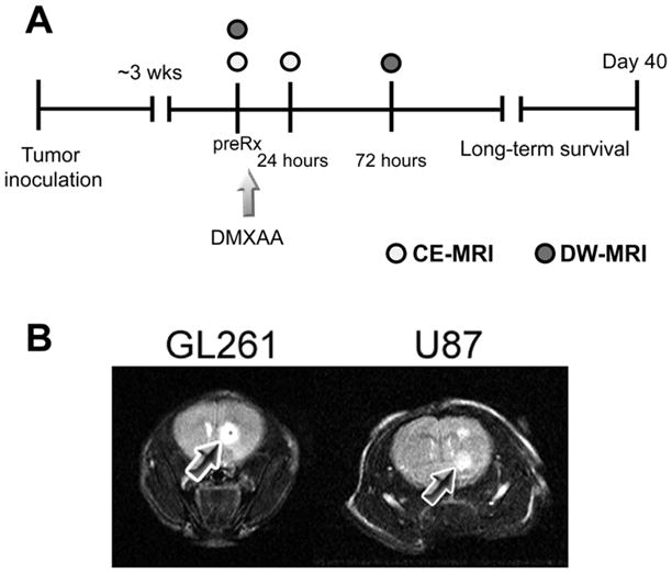
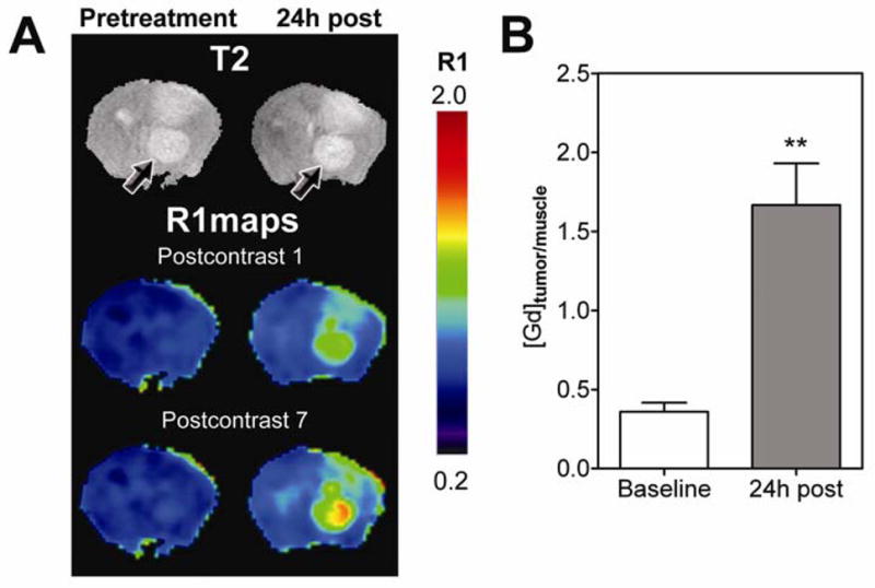
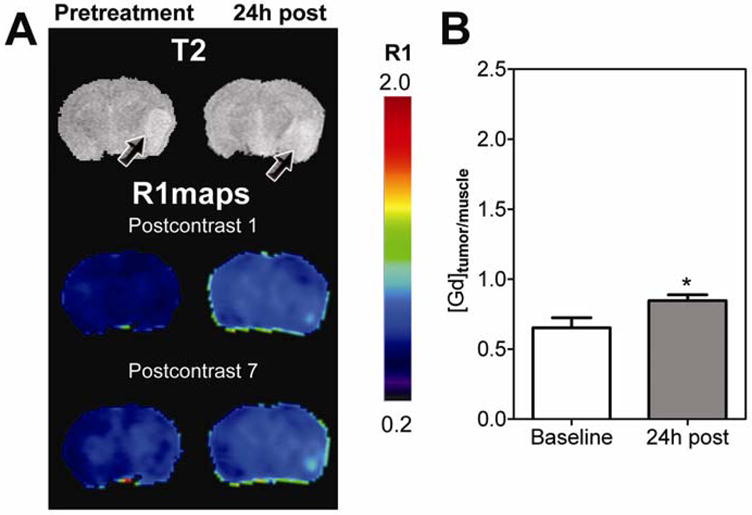
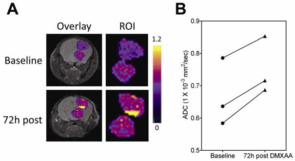
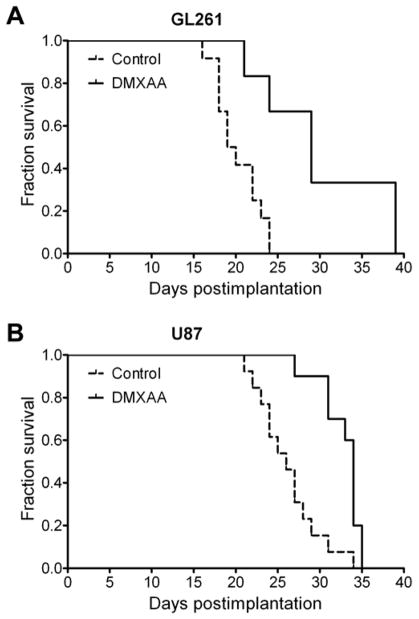
The well-vascularized nature of gliomas has generated a lot of interest in antiangiogenic therapies. However, the potential of vascular disrupting agents (VDAs) against gliomas has not been investigated extensively. In this study, we examined the in vivo efficacy of the tumor-VDA 5,6-dimethylxanthenone-4-acetic acid (DMXAA) against gliomas. Contrast-enhanced magnetic resonance imaging (MRI) and diffusion-weighted MRI were used to characterize the vascular and cellular responses of GL261 and U87 gliomas to DMXAA treatment. Therapeutic efficacy was assessed by Kaplan-Meier survival analysis. Before VDA treatment, minimal enhancement was detected within the tumor in both models. Longitudinal relaxation rate (R1=1/T1) maps acquired 24 h after treatment showed marked extravasation and accumulation of the contrast agent in the tumor indicative of treatment-induced vascular disruption. Normalized change in relaxation rate (DeltaR1) values of the tumor showed a significant increase (P<0.01 GL261; P<0.05 U87) after therapy compared with baseline estimates. Mean apparent diffusion coefficient (ADC) values were significantly increased (P=0.015) 72 h after therapy in GL261 but not in U87 gliomas. Vascular disrupting agent therapy resulted in a significant (P<0.01) increase in median survival in both models evaluated. The results highlight the potential of VDAs against gliomas and the utility of MRI in the assessment of glioma response to VDA therapy.
Figures
Similar articles
-
Bioluminescence and MR Imaging of the Safety and Efficacy of Vascular Disruption in Gliomas.Mol Imaging Biol. 2016 Dec;18(6):860-869. doi: 10.1007/s11307-016-0963-8. Mol Imaging Biol. 2016. PMID: 27160251
-
Efficacy against subcutaneous or intracranial murine GL261 gliomas in relation to the concentration of the vascular-disrupting agent, 5,6-dimethylxanthenone-4-acetic acid (DMXAA), in the brain and plasma.Cancer Chemother Pharmacol. 2014 Mar;73(3):639-49. doi: 10.1007/s00280-014-2395-y. Epub 2014 Jan 30. Cancer Chemother Pharmacol. 2014. PMID: 24477604
-
Assessment of the early effects of 5,6-dimethylxanthenone-4-acetic acid using macromolecular contrast media-enhanced magnetic resonance imaging: ectopic versus orthotopic tumors.Int J Radiat Oncol Biol Phys. 2008 Nov 15;72(4):1198-207. doi: 10.1016/j.ijrobp.2008.07.043. Int J Radiat Oncol Biol Phys. 2008. PMID: 18954713 Free PMC article.
-
Current standards and new concepts in MRI and PET response assessment of antiangiogenic therapies in high-grade glioma patients.Neuro Oncol. 2015 Jun;17(6):784-800. doi: 10.1093/neuonc/nou322. Epub 2014 Dec 27. Neuro Oncol. 2015. PMID: 25543124 Free PMC article. Review.
-
Imaging biomarkers guided anti-angiogenic therapy for malignant gliomas.Neuroimage Clin. 2018 Jul 5;20:51-60. doi: 10.1016/j.nicl.2018.07.001. eCollection 2018. Neuroimage Clin. 2018. PMID: 30069427 Free PMC article. Review.
Cited by
-
Non-Invasive Evaluation of Acute Effects of Tubulin Binding Agents: A Review of Imaging Vascular Disruption in Tumors.Molecules. 2021 Apr 27;26(9):2551. doi: 10.3390/molecules26092551. Molecules. 2021. PMID: 33925707 Free PMC article. Review.
-
The influence of the combined treatment with Vadimezan (ASA404) and taxol on the growth of U251 glioblastoma xenografts.BMC Cancer. 2012 Jun 13;12:242. doi: 10.1186/1471-2407-12-242. BMC Cancer. 2012. PMID: 22695475 Free PMC article.
-
A perspective on vascular disrupting agents that interact with tubulin: preclinical tumor imaging and biological assessment.Integr Biol (Camb). 2011 Apr;3(4):375-87. doi: 10.1039/c0ib00135j. Epub 2011 Feb 14. Integr Biol (Camb). 2011. PMID: 21321746 Free PMC article.
-
Impact of Blood-Brain Barrier to Delivering a Vascular-Disrupting Agent: Predictive Role of Multiparametric MRI in Rodent Craniofacial Metastasis Models.Cancers (Basel). 2022 Nov 26;14(23):5826. doi: 10.3390/cancers14235826. Cancers (Basel). 2022. PMID: 36497308 Free PMC article.
-
Antitumor cytotoxic T-cell response induced by a survivin peptide mimic.Cancer Immunol Immunother. 2010 Aug;59(8):1211-21. doi: 10.1007/s00262-010-0845-x. Epub 2010 Apr 27. Cancer Immunol Immunother. 2010. PMID: 20422411 Free PMC article.
References
-
- Abdulrauf SI, Edvardsen K, Ho KL, Yang XY, Rock JP, Rosenblum ML. Vascular endothelial growth factor expression and vascular density as prognostic markers of survival in patients with low-grade astrocytoma. J Neurosurg. 1998;88:513–20. - PubMed
-
- Akella NS, Twieg DB, Mikkelsen T, Hochberg FH, Grossman S, Cloud GA, Nabors LB. Assessment of brain tumor angiogenesis inhibitors using perfusion magnetic resonance imaging: quality and analysis results of a phase I trial. J Magn Reson Imaging. 2004;20:913–22. - PubMed
-
- Baguley BC, Wilson WR. Potential of DMXAA combination therapy for solid tumors. Expert Rev Anticancer Ther. 2002;2:593–603. - PubMed
-
- Barrett T, Kobayashi H, Brechbiel M, Choyke Macromolecular MRI contrast agents for imaging tumor angiogenesis. Eur J Radiol. 2006;60:353–66. - PubMed
-
- Barth RF, Yang W, Rotaru JH, Moeschberger ML, Boesel CP, Soloway AH, Joel DD, Nawrocky MM, Ono K, Goodman JH. Boron neutron capture therapy of brain tumors: enhanced survival and cure following blood-brain barrier disruption and intracarotid injection of sodium borocaptate and borophenylalanine. Int J Radiat Oncol Biol Phys. 2000;47:209–18. - PubMed
Publication types
MeSH terms
Substances
Grants and funding
LinkOut - more resources
Full Text Sources
Medical
