. 2009;154(6):945-57.
doi: 10.1007/s00705-009-0393-x. Epub 2009 May 21.

Functional significance of the hemadsorption activity of influenza virus neuraminidase and its alteration in pandemic viruses

Affiliations

Functional significance of the hemadsorption activity of influenza virus neuraminidase and its alteration in pandemic viruses

Jennifer Uhlendorff et al. Arch Virol. 2009.

Abstract

Human influenza viruses derive their genes from avian viruses. The neuraminidase (NA) of the avian viruses has, in addition to the catalytic site, a separate sialic acid binding site (hemadsorption site) that is not present in human viruses. The biological significance of the NA hemadsorption activity in avian influenza viruses remained elusive. A sequence database analysis revealed that the NAs of the majority of human H2N2 viruses isolated during the influenza pandemic of 1957 differ from their putative avian precursor by amino acid substitutions in the hemadsorption site. We found that the NA of a representative pandemic virus A/Singapore/1/57 (H2N2) lacks hemadsorption activity and that a single reversion to the avian-virus-like sequence (N367S) restores hemadsorption. Using this hemadsorption-positive NA, we generated three NA variants with substitutions S370L, N400S and W403R that have been found in the hemadsorption site of human H2N2 viruses. Each substitution abolished hemadsorption activity. Although, there was no correlation between hemadsorption activity of the NA variants and their enzymatic activity with respect to monovalent substrates, all four hemadsorption-negative NAs desialylated macromolecular substrates significantly slower than did the hemadsorption-positive counterpart. The NA of the 1918 pandemic virus A/Brevig Mission/1/18 (H1N1) also differed from avian N1 NAs by reduced hemadsorption activity and less efficient hydrolysis of macromolecular substrates. Our data indicate that the hemadsorption site serves to enhance the catalytic efficiency of NA and they suggest that, in addition to changes in the receptor-binding specificity of the hemagglutinin, alterations of the NA are needed for the emergence of pandemic influenza viruses.


Figures

Fig. 1
Fig. 1
Hemadsorption site of the NA and its alteration in the H2N2 pandemic viruses. a Molecular surface of N9 NA tetramer with sialic acids bound to the catalytic (green) and hemadsorption (yellow) sites (1MWE, [48]). b Positions of the three peptide loops forming the hemadsorption site are indicated. The side chains of amino acids contacting sialic acid are shown and labelled using the N2 numbering system. The carbon atoms of the protein and sialic acid are coloured yellow and grey, respectively. Nitrogens are blue and oxygens are red. Dashed lines depict polar contacts. The models in Figs. 1 and 5 were generated using DeLano Scientific PyMOL release 0.99 (DeLano, W.L. The PyMOL Molecular Graphics System, http://pymol.sourceforge.net). c Amino acid substitutions in the NA hemadsorption site of human viruses with respect to the closely related avian virus A/Duck/Hong Kong/7/75 (H3N2). All non-redundant NA sequences of H2N2 viruses from 1957 to 1958 and few representative sequences of the viruses isolated after 1958 are shown. Amino acids contacting sialic acid in the hemadsorption site are highlighted. The minimum evolution phylogenetic tree was build for nucleotide sequences using MEGA4 software [44]
Fig. 2
Fig. 2
Hemadsorption activity of N2 neuraminidases. Cos-7 cells were transfected with pCAGGS-NA plasmids coding for either SG/57 NA or its variants with indicated amino acid substitutions. Empty pCAGGS plasmid was used as a control (mock). Forty-eight hours post transfection, the cells were incubated with chicken (left panel) and human (right panel) erythrocytes, which are commonly used to characterize binding specificity of influenza viruses. The cells were stained and photographed using Olympus IMT-2 microscope equipped with Nikon DS-2Mv camera. Objective, 20×. Data are representative of five or more independent experiments
Fig. 3
Fig. 3
Neuraminidase activity of the recombinant viruses rgHAD− and rgHAD+. Viral suspensions were adjusted to the same NA content (a) and compared for their enzymatic activity with respect to soluble substrates (b) and cells (c). Data are representative of five or more independent experiments. a Western blot analysis of rgHAD− (lane 1) and rgHAD+ (lane 2) using anti-H9N2 influenza virus antibodies. Lysates of 293T cells transfected with pCAGGS plasmids expressing M1 protein of A/Hong Kong/1/68 (lane 3) and NA of A/Singapore/1/57 (lane 4) were included as controls. b rgHAD− (blue) and rgHAD+ (red) were incubated with substrates containing 0.1 mM sialic acid at 37°C, and concentrations of released sialic acid were determined at different time points. To account for substrate-specific differences in viral neuraminidase activity, 20-fold lower virus concentration was used in the case of 3SLN, 3SL-PAA and MU-NANA than in the case of other four substrates. Mean values and SD of two replicate samples in one experiment are shown. c Serial twofold dilutions of the viruses were incubated with equal volumes of 0.5% human erythrocytes at 4°C for 1 h (top panel), followed by incubation at 37°C for 8 h (bottom panel)
Fig. 4
Fig. 4
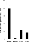
NA hemadsorption to native and modified erythrocytes. Cos-7 cells expressing the hemadsorption-positive NA variant N367S were probed with native human erythrocytes, erythrocytes desialylated with V. cholerae sialidase, and erythrocytes resialylated using α2-3- and α2-6-sialyltransferases. Data show mean numbers of red blood cells attached per ten Cos-7 cells and are representative of three independent experiments
Fig. 5
Fig. 5
Comparison of the structures of the NA hemadsorption site of the N9 NA with bound sialic acid [48] and of the HA receptor-binding site of A/duck/Ukraine/63 (H3N8) with bound Neu5Acα2-3Gal disaccharide [15]. The ligands are shown as stick models. Hemadsorption site (yellow) and receptor-binding site (green) are superimposed over the bound sialic acid residues. Positions of peptide loops forming the sites are indicated
Fig. 6
Fig. 6
Hemadsorption activity of N1 neuraminidases. Cos-7 cells were transfected with pCAGGS-NA plasmids coding for the neuraminidases of A/Brevig Mission/1/18 (H1N1), A/chicken/FPV/Rostock/34 (H7N1) and A/Ostrich/Italy/984/99 (H7N1). pCAGGS plasmids expressing N2 NAs of SG/57 and its hemadsorption-positive mutant (N367S) were used as negative and positive controls, respectively. Forty-eight hours post transfection, the cells were probed with chicken (closed bars) and human (hatched bars) erythrocytes. Data are mean values and SD of replicate samples in one experiment representative of five independent experiments. The values of hemadsorption activity of the 1918 NA differ from corresponding values of each other NAs tested (P < 0.05, unpaired two-sided t test)

References

    1. {'text': '', 'ref_index': 1, 'ids': [{'type': 'DOI', 'value': '10.1016/j.virol.2006.10.037', 'is_inner': False, 'url': 'https://doi.org/10.1016/j.virol.2006.10.037'}, {'type': 'PMC', 'value': 'PMC2735206', 'is_inner': False, 'url': 'https://pmc.ncbi.nlm.nih.gov/articles/PMC2735206/'}, {'type': 'PubMed', 'value': '17157891', 'is_inner': True, 'url': 'https://pubmed.ncbi.nlm.nih.gov/17157891/'}]}
    2. Aamir UB, Wernery U, Ilyushina N, Webster RG (2007) Characterization of avian H9N2 influenza viruses from United Arab Emirates 2000 to 2003. Virology 361:45–55 - PMC - PubMed
    1. {'text': '', 'ref_index': 1, 'ids': [{'type': 'DOI', 'value': '10.1002/prot.340060402', 'is_inner': False, 'url': 'https://doi.org/10.1002/prot.340060402'}, {'type': 'PubMed', 'value': '2482974', 'is_inner': True, 'url': 'https://pubmed.ncbi.nlm.nih.gov/2482974/'}]}
    2. Air GM, Laver WG (1989) The neuraminidase of influenza virus. Proteins 6:341–356 - PubMed
    1. {'text': '', 'ref_index': 1, 'ids': [{'type': 'DOI', 'value': '10.1006/viro.1995.1401', 'is_inner': False, 'url': 'https://doi.org/10.1006/viro.1995.1401'}, {'type': 'PubMed', 'value': '7645221', 'is_inner': True, 'url': 'https://pubmed.ncbi.nlm.nih.gov/7645221/'}]}
    2. Air GM, Laver WG (1995) Red cells bound to influenza virus N9 neuraminidase are not released by the N9 neuraminidase activity. Virology 211:278–284 - PubMed
    1. {'text': '', 'ref_index': 1, 'ids': [{'type': 'DOI', 'value': '10.1002/prot.340020205', 'is_inner': False, 'url': 'https://doi.org/10.1002/prot.340020205'}, {'type': 'PubMed', 'value': '3447170', 'is_inner': True, 'url': 'https://pubmed.ncbi.nlm.nih.gov/3447170/'}]}
    2. Baker AT, Varghese JN, Laver WG, Air GM, Colman PM (1987) Three-dimensional structure of neuraminidase of subtype N9 from an avian influenza virus. Proteins 2:111–117 - PubMed
    1. {'text': '', 'ref_index': 1, 'ids': [{'type': 'DOI', 'value': '10.1016/0042-6822(91)90003-T', 'is_inner': False, 'url': 'https://doi.org/10.1016/0042-6822(91)90003-t'}, {'type': 'PubMed', 'value': '1984642', 'is_inner': True, 'url': 'https://pubmed.ncbi.nlm.nih.gov/1984642/'}]}
    2. Baum LG, Paulson JC (1991) The N2 neuraminidase of human influenza virus has acquired a substrate specificity complementary to the hemagglutinin receptor specificity. Virology 180:10–15 - PubMed

Publication types

MeSH terms

LinkOut - more resources