

Pancreatic serous cystadenocarcinoma: a case report and review of the literature
- PMID: 19459016
- PMCID: PMC2759006
- DOI: 10.1007/s11605-009-0926-3
Pancreatic serous cystadenocarcinoma: a case report and review of the literature
Abstract
Background: Serous cystic neoplasms of the pancreas are benign lesions with little chance for malignant degeneration. We report a case of malignant serous cystadenocarcinoma of the pancreas and review the literature.
Methods: Structured review of the literature was performed using PubMed and MEDLINE searches, and cases of serous cystadenocarcinoma of the pancreas were compiled.
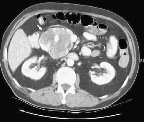
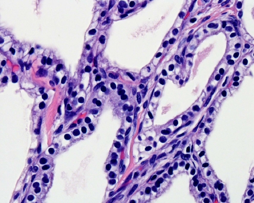
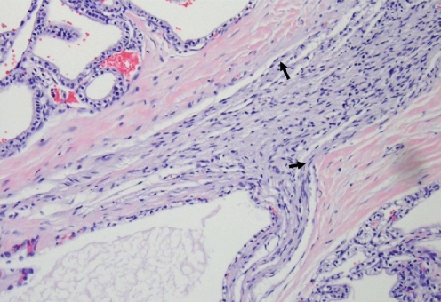
Results: A 70-year-old man diagnosed with a serous cystadenoma was managed expectantly until he became symptomatic, and studies revealed an increase in the size of the lesion as well as duodenal invasion. The patient underwent a pancreaticoduodenectomy, and histopathological examination revealed a locally invasive cystadenocarcinoma without metastatic disease. Seven years later, the patient remains disease-free. Review of the literature identified 25 cases of serous cystadenocarcinoma published to date. The mean age at diagnosis is 68 +/- 2 years (range, 52 to 81), and women are affected more commonly (2:1).
Conclusions: We conclude that there is a small but finite risk of malignancy for serous cystic neoplasms of the pancreas. The clinician should bear this in mind when faced with decisions regarding patient management. Prognosis is excellent with multiple reports of long-term survival even in the face of metastatic disease.
Figures


References
-
- {'text': '', 'ref_index': 1, 'ids': [{'type': 'DOI', 'value': '10.1097/00000658-196506000-00005', 'is_inner': False, 'url': 'https://doi.org/10.1097/00000658-196506000-00005'}, {'type': 'PMC', 'value': 'PMC1409085', 'is_inner': False, 'url': 'https://pmc.ncbi.nlm.nih.gov/articles/PMC1409085/'}, {'type': 'PubMed', 'value': '14295937', 'is_inner': True, 'url': 'https://pubmed.ncbi.nlm.nih.gov/14295937/'}]}
- Becker WF, Welsh A, Pratt HS. Cystadenoma and cystadenocarcinoma of the pancreas. Ann Surg 1965;161:845–860. doi:10.1097/00000658-196506000-00005. - PMC - PubMed
-
- None
- Klöppel G, Solcia E, Longnecker DS, Capella C, Sobin LH. Histological typing of tumors of the exocrine pancreas, 2nd edition. WHO international histological classification of tumors. Berlin: Springer, 1996.
-
- {'text': '', 'ref_index': 1, 'ids': [{'type': 'PubMed', 'value': '665578', 'is_inner': True, 'url': 'https://pubmed.ncbi.nlm.nih.gov/665578/'}]}
- Compagno J, Oertel JE. Mucinous cystic neoplasms of the pancreas with overt and latent malignancy (cystadenocarcinoma and cystadenoma): A clinicopathological study of 41 cases. Am J Clin Pathol 1978;69:573–580. - PubMed
-
- {'text': '', 'ref_index': 1, 'ids': [{'type': 'PubMed', 'value': '637043', 'is_inner': True, 'url': 'https://pubmed.ncbi.nlm.nih.gov/637043/'}]}
- Compagno J, Oertel JE. Microcystic adenomas of the pancreas (glycogen-rich cystadenomas). Am J Clin Pathol 1978;69:289–298. - PubMed
-
- {'text': '', 'ref_index': 1, 'ids': [{'type': 'PubMed', 'value': '2909198', 'is_inner': True, 'url': 'https://pubmed.ncbi.nlm.nih.gov/2909198/'}]}
- George DH, Murphy F, Michalski R, Ulmer BG. Serous cystadenocarcinoma of the pancreas: A new entity? Am J Surg Pathol 1989;13:61–66. doi:10.1097/00000478-198901000-00009. - PubMed
