Review
. 2009 Aug;66(8):606-17.
doi: 10.1002/cm.20379.

Review of the mechanism of processive actin filament elongation by formins

Affiliations
Review

Review of the mechanism of processive actin filament elongation by formins

Aditya S Paul et al. Cell Motil Cytoskeleton. 2009 Aug.

Abstract

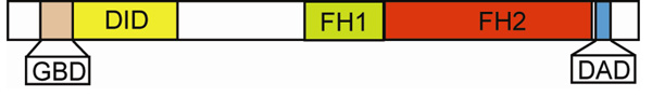
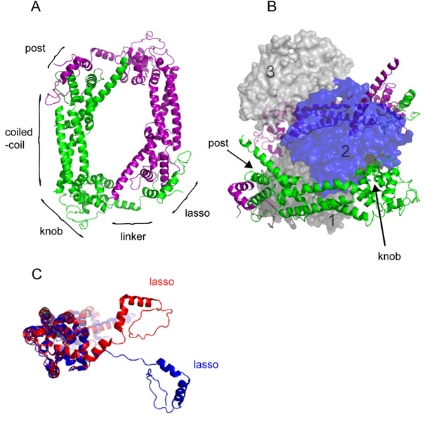
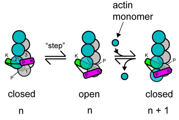
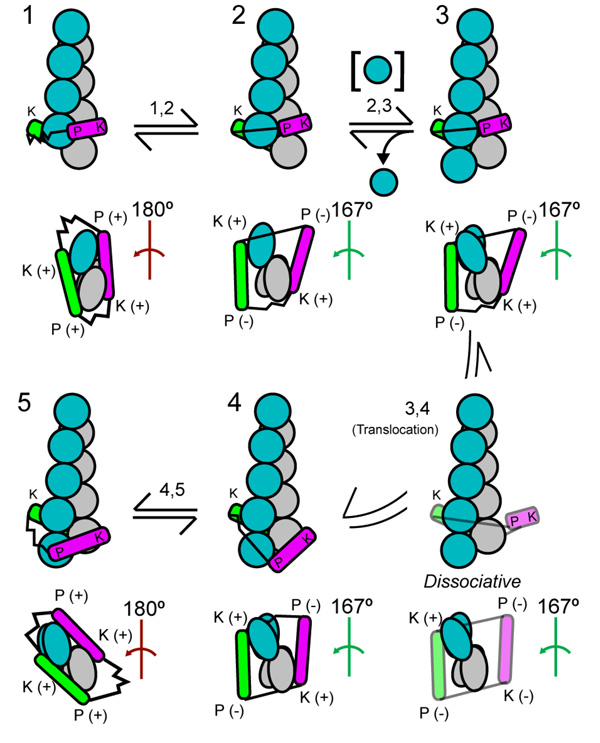
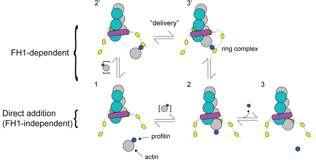
We review recent structural and biophysical studies of the mechanism of action of formins, proteins that direct the assembly of unbranched actin filaments for cytokinetic contractile rings and other cellular structures. Formins use free actin monomers to nucleate filaments and then remain bound to the barbed ends of these filaments as they elongate. In addition to variable regulatory domains, formins typically have formin homology 1 (FH1) and formin homology 2 (FH2) domains. FH1 domains have multiple binding sites for profilin, an abundant actin monomer binding protein. FH2 homodimers encircle the barbed end of a filament. Most FH2 domains inhibit actin filament elongation, but FH1 domains concentrate multiple profilin-actin complexes near the end of the filament. FH1 domains transfer actin very rapidly onto the barbed end of the filament, allowing elongation at rates that exceed the rate of elongation by the addition of free actin monomers diffusing in solution. Binding of actin to the end of the filament provides the energy for the highly processive movement of the FH2 as a filament adds thousands of actin subunits. These biophysical insights provide the context to understand how formins contribute to actin assembly in cells. Cell Motil. Cytoskeleton 2009. (c) 2009 Wiley-Liss, Inc.


Figures

Figure 1
Figure 1. Domain map of the formin mDia1
The arrangement of the GTPase-binding domain (GBD), the Diaphanous-Inhibitory Domain (DID), the Formin-homology (FH1) Domain, the Formin-homology (FH2) Domain, and Diaphanous Autoregulatory Domain (DAD), are delineated at their approximate, relative scales according to primary sequence of the full-length mDia1 formin molecule (Higgs 2005).
Figure 2
Figure 2. The structure of the Bni1p-FH2 domain
(A) Ribbon diagram of the crystal structure of the head to tail homodimer of FH2 domains of Bni1p (residues 1350–1760) (Xu et al. 2004) (PDB Accession Code 1UX5). The two subunits are shown in green and purple. Labels indicate the approximate positions of the lasso, flexible linker, knob, coiled-coil, and post regions of the green subunit. (B) Crystal structure of the complex of Bni1p-FH2 (residues 1350–1760) with muscle actin (Otomo et al. 2005b) (PDB Accession Code 1Y64). Three contiguous actin subunits along the filament-like polymer are shown as space-filling representation in shades of gray and blue and numbered 1 to 3 from the barbed end. Ribbon diagrams of two FH2 subunits are colored as in (A). A continuous chain of FH2 domains wraps around the actin polymer with the lasso of one FH2 subunit joined to the post of the next. For clarity the density for the linker (residues 1401–1417) is omitted. This view shows the knob of the green FH2 subunit bound in the groove between subdomains 1 and 3 of actin subunit 2, as well as the post site of the green FH2 subunit bound to subdomain 1 of actin subunit 2. The partial transparency of the actin subunits reveals the symmetrical attachments of the purple FH2 subunit to actin subunits 2 and 3. (C) Comparison of the FH2 subunits from (A) and (B). When the knob and post regions of Bni1p-FH2 (residues 1418–1760) from the homodimer (Xu et al. 2004) (red) and the cocrystal with actin (Otomo et al. 2005b) (blue) were overlaid with PYMOL using the “align” function, the positions of the lasso-linkers diverge substantially with the linker more extended in complex with actin. All images were rendered with PYMOL (Delano Scientific).
Figure 3
Figure 3. Two-state “stair-stepping” model for processive association of the formin-FH2 dimer with a growing barbed end
The schematic depicts addition of one subunit (from “n” to “n+1”) onto barbed end associated with a FH2 dimer, where the formin equilibrates between a closed state that prevents subunit addition and an open state that allows addition (Otomo et al. 2005b; Xu et al. 2004). The subunits of the two long-pitch strands of the actin filament are shown as blue or silver spheres numbered 1, 2 and 3 from the barbed end. The leading subunit of the FH2 dimer is green and the trailing subunit purple. In the closed state, the FH2 dimer is engaged at both of its knob (K) and post (P) sites to the three terminal subunits. The leading FH2 subunit binds to actin subunits 1 and 2 and the trailing subunit binds to actin subunits 2 and 3 in the closed state. To enter the open state, the trailing subunit disengages both its knob and post sites, translocates or “steps” in the barbed end direction, and reattaches only its knob to the terminal barbed end subunit. In this open state, this FH2 subunit’s post site is exposed to solution. An actin monomer in solution binds to this post site and the two terminal actin subunits to complete the cycle of subunit addition and reestablish the closed state.
Figure 4
Figure 4. The “stepping second” hypothesis for actin subunit addition to a barbed end associated with a formin FH2 domain
The drawing gives five steps (1–5) and transitions in a hypothetical mechanical cycle of actin subunit addition coupled to translocation of a formin FH2 dimer (green and magenta). States 5 and 4 are equivalent to states 1 and 2 but the filament is one subunit longer. The upper images for each state show a side view with the barbed end down. The lower images are views of the barbed end. The actin subunits are grey along one long pitch strand and blue along the other strand. The short-pitch helical twist of the 3 terminal barbed end subunits is either 180° as found in cocrystals of Bni1pFH2 with actin or the 167° as in the core of actin filaments. The red angle symbol indicates closed 180° conformations that do not accept subunit addition. The green angle symbol indicates open 167° conformations that permit subunit addition. Each FH2 subunit has two sites that can interact with actin: the knob (K) and post (P). Sites engaged with the filament are labeled (+). Sites dissociated from the filament are labeled (−). The flexible linkers between the two FH2 subunits are depicted as either stretched or relaxed springs. States 1 and 2 (as 5 and 4) are rapid equilibria between the open and closes states. A new actin subunit adds to open state 2 to form intermediate state 3. The leading FH2 subunit steps onto the new terminal subunit to complete the cycle. (From Paul and Pollard, Paul and Pollard 2008)
Figure 5
Figure 5. Comparison of FH1-dependent and independent pathways of actin subunit addition
Through the process of subunit addition, each state of the formin-FH1FH2-associated end is denoted by a different number. The actin filament and formin dimer subunits are colored as in Figure 3. Each FH1 domain has multiple polyproline tracks (yellow ovals). Profilin (small blue circle) binds to an actin monomer in solution (large gray circle). To complete one cycle of subunit addition, the profilin-actin complex may add onto the formin-FH1FH2-associated end via the FH1-independent pathway (1-2-3) or the FH1-dependent pathway (1-2’-3’-2-3) (Vavylonis et al. 2006). The rapid delivery step by which FH1-bound-profilin-actin is transferred directly to the FH2-associated barbed end and the resultant “ring complex” (Vavylonis et al. 2006) are labeled.

References

    1. Alberts AS. Identification of a carboxyl-terminal diaphanous-related formin homology protein autoregulatory domain. J Biol Chem. 2001;276(4):2824–2830. - PubMed
    1. Andrianantoandro E, Pollard TD. Mechanism of actin filament turnover by severing and nucleation at different concentrations of ADF/cofilin. Mol Cell. 2006;24(1):13–23. - PubMed
    1. Archer SJ, Vinson VK, Pollard TD, Torchia DA. Elucidation of the poly-L-proline binding site in Acanthamoeba profilin-I by NMR spectroscopy. FEBS Lett. 1994;337:145–151. - PubMed
    1. Baker JE, Krementsova EB, Kennedy GG, Armstrong A, Trybus KM, Warshaw DM. Myosin V processivity: multiple kinetic pathways for head-to-head coordination. Proc Natl Acad Sci USA. 2004;101:5542–5546. - PMC - PubMed
    1. Blanchoin L, Pollard TD. Hydrolysis of ATP by polymerized actin depends on the bound divalent cation but not profilin. Biochemistry. 2002;41(2):597–602. - PubMed

Publication types

LinkOut - more resources