. 2009 May;276(10):2875-90.
doi: 10.1111/j.1742-4658.2009.07011.x.

Functional aspects of the solution structure and dynamics of PAF--a highly-stable antifungal protein from Penicillium chrysogenum

Affiliations

Functional aspects of the solution structure and dynamics of PAF--a highly-stable antifungal protein from Penicillium chrysogenum

Gyula Batta et al. FEBS J. 2009 May.

Abstract

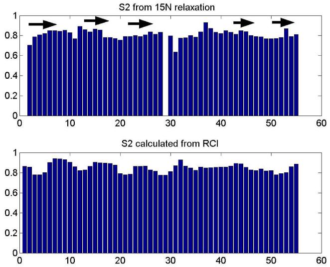
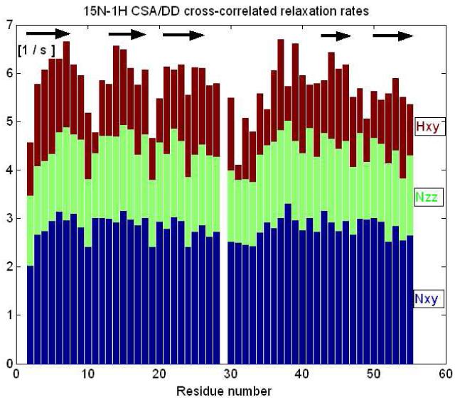
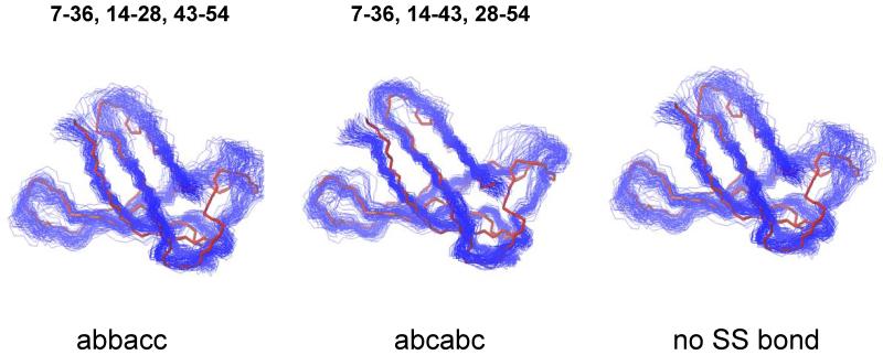
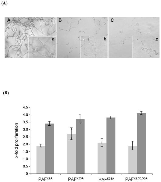
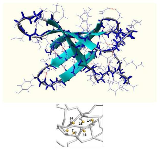
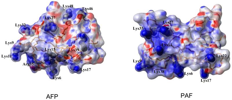
Penicillium antifungal protein (PAF) is a promising antimycotic without toxic effects on mammalian cells and therefore may represent a drug candidate against the often lethal Aspergillus infections that occur in humans. The pathogenesis of PAF on sensitive fungi involves G-protein coupled signalling followed by apoptosis. In the present study, the solution structure of this small, cationic, antifungal protein from Penicillium chrysogenum is determined by NMR. We demonstrate that PAF belongs to the structural classification of proteins fold class of its closest homologue antifungal protein from Aspergillus giganteus. PAF comprises five beta-strands forming two orthogonally packed beta-sheets that share a common interface. The ambiguity in the assignment of two disulfide bonds out of three was investigated by NMR dynamics, together with restrained molecular dynamics calculations. The clue could not be resolved: the two ensembles with different disulfide patterns and the one with no S-S bond exhibit essentially the same fold. (15)N relaxation dispersion and interference experiments did not reveal disulfide bond rearrangements via slow exchange. The measured order parameters and the 3.0 ns correlation time are appropriate for a compact monomeric protein of this size. Using site-directed mutagenesis, we demonstrate that the highly-conserved and positively-charged lysine-rich surface region enhances the toxicity of PAF. However, the binding capability of the oligosaccharide/oligonucleotide binding fold is reduced in PAF compared to antifungal protein as a result of less solvent-exposed aromatic regions, thus explaining the absence of chitobiose binding. The present study lends further support to the understanding of the documented substantial differences between the mode of action of two highly homologous antifungal proteins.


Figures

Fig. 1
Fig. 1
Microtiterplate activity assay for the determination of the growth of A. niger in the presence of PAF that had been exposed to various test conditions. 104 conidia/ml were incubated with 1-5 μg/ml PAF for 24 h at 30°C. (A) PAF was reduced by DTT as described in Material and Methods. (B) PAF was exposed to 60°C, 80°C, 90°C and 100°C for 10, 30, 60 min, respectively. (C) PAF was digested with proteinase K for 3, 9 and 24 h or with pronase for 3, 9, and 12 h. Please note, that the asterisk in (C) indicates different exposure times of PAF with pronase (12 h instead of 24 h). Values represent the precent growth (%) of A. niger in the presence of PAF that had been exposed to various test conditions compared to A. niger left untreated (=100%).
Fig. 2
Fig. 2
Sequence alignment of PAF and AFP with three highly conserved regions marked in yellow that are putatively assigned to chitin binding (3-9), DNA binding (12-17) domains and cation channel forming (34-39) capabilities. Arrows and S labels stand for β strands.
Fig. 3
Fig. 3
Super secondary structure of PAF
Fig. 4
Fig. 4
Deuteration rates measured for amide NH groups in PAF (note the logarithmic scale). Missing bars represent fast deuteration rates, i.e. those NH signals disappeared within 10 minutes. Slow deuteration of amides protons correlates with solvent protection in β-sheet regions.
Fig. 5
Fig. 5
S2 order parameters reflecting internal mobility of the NH residues obtained from the Lipari-Szabo analysis of 15N T1, T2 and NOE relaxation parameters. Slightly enhanced mobility is clearly detected at the N-terminus and in the loop regions as shown by the dips of the bar plot. For comparison, the S2 values calculated from the assigned chemical shifts is shown at the bottom using the RCI index and the program of http://wishart.biology.ualberta.ca/rci/cgi-bin/rci_cgi_1_e.py. The average of S2exp/S2RCI = 0.96 ± 0.07. Residue 29 is proline and consequently not shown in experimental data.
Fig. 6
Fig. 6
Different CSA/DD relaxation interference rates displayed as a pile-up bar graph. Instead of extracting site specific 15N chemical-shift anisotropies from all relaxation data, this kind of straight visualisation of 1H- 15N CSA/ DD transversal cross-correlated relaxation rates (Hxy) is sensitive to secondary structure elements.
Figure 7
Figure 7
Final MUMO ensembles (80 conformers) of PAF calculated with different disulfide pairings labelled as abbacc, abcabc and no SS bond. The average NMR structure (red line) with no disulfide bonds is overlayed with the ensembles.
Fig. 8
Fig. 8
Analysis of the antifungal activity of mutated PAF protein versions on A. niger. (A) Microscopic analysis of A. niger exposed to 100 μg/ml PAF for 24 h. The recombinant mPAF protein (panel C) exhibited comparable growth inhibition potency to the native PAF (panel B). In panel A hyphae of the untreated control are shown. Panels A-C are microscopic overviews (×20), panels a-c are details of A-C (×63). (B) The increase in proliferation of A. niger when exposed to mutated PAF protein versions was correlated to the proliferation in the presence of recombinant mPAF at the corresponding protein concentrations of 5 μg/ml (light grey) and 100 μg/ml (dark grey). The proliferation of the PAF-unexposed A. niger control cells was 2.4(±0.2)-fold and 7.4(±1.1)-fold compared to the growth of the samples treated with recombinant mPAF at the respective concentrations of 5 μg/ml and 100 μg/ml.
Fig. 9
Fig. 9
Ribbon diagram of the mean PAF structure without disulfide bond constraints. The congested hydrophobic core with the cysteines labelled is shown in the excerpt.
Fig. 10
Fig. 10
The electrostatic surface potenctial of AFP (left) and PAF (right) structures representing the orientation of the side chain Lysine resiues, Lys9, Lys35, Lys 38 of PAF and Lys9, Lys32 and Arg35 of AFP are the corresponding conservative mutated lysines.

References

    1. Marx F. Small, basic antifungal proteins secreted from filamentous ascomycetes: a comparative study regarding expression, structure, function and potential application. Applied Microbiology and Biotechnology. 2004;65:133–142. - PubMed
    1. Geisen R. P-nalgiovense carries a gene which is homologous to the paf gene of P-chrysogenum which codes for an antifungal peptide. International Journal of Food Microbiology. 2000;62:95–101. - PubMed
    1. Kaiserer L, Oberparleiter C, Weiler-Gorz R, Burgstaller W, Leiter E, Marx F. Characterization of the Penicillium chrysogenum antifungal protein PAF. Archives of Microbiology. 2003;180:204–210. - PubMed
    1. Lee DG, Shin SY, Maeng CY, Jin ZZ, Kim KL, Hahm KS. Isolation and characterization of a novel antifungal peptide from Aspergillus niger. Biochemical and Biophysical Research Communications. 1999;263:646–651. - PubMed
    1. Marx F, Haas H, Reindl M, Stoffler G, Lottspeich F, Redl B. Cloning, structural organization and regulation of expression of the Penicillium chrysogenum paf gene encoding an abundantly secreted protein with antifungal activity. Gene. 1995;167:167–171. - PubMed

Publication types

MeSH terms

Associated data