Comparative Study
. 2009 Aug;47(2):68-77.
doi: 10.1016/j.micpath.2009.04.015. Epub 2009 May 19.

The structurally similar, penta-acylated lipopolysaccharides of Porphyromonas gingivalis and Bacteroides elicit strikingly different innate immune responses

Affiliations
Comparative Study

The structurally similar, penta-acylated lipopolysaccharides of Porphyromonas gingivalis and Bacteroides elicit strikingly different innate immune responses

Alex B Berezow et al. Microb Pathog. 2009 Aug.

Abstract

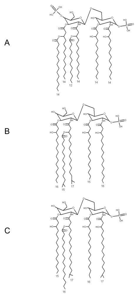
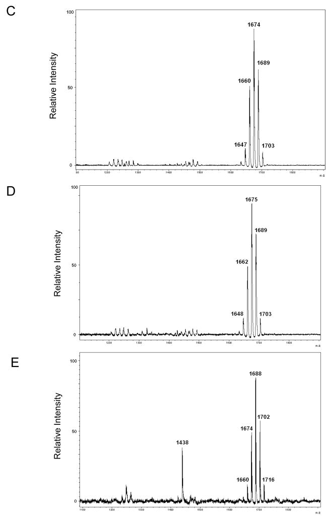
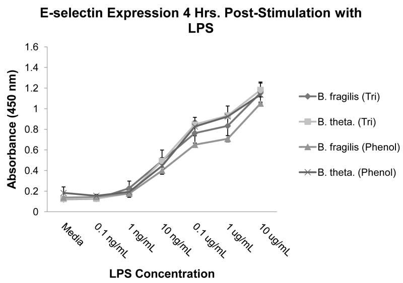
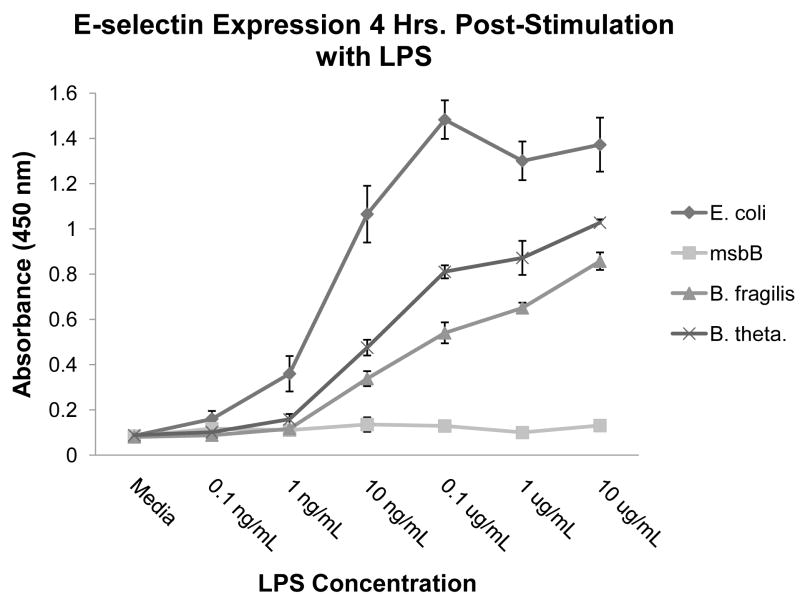
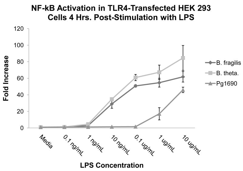
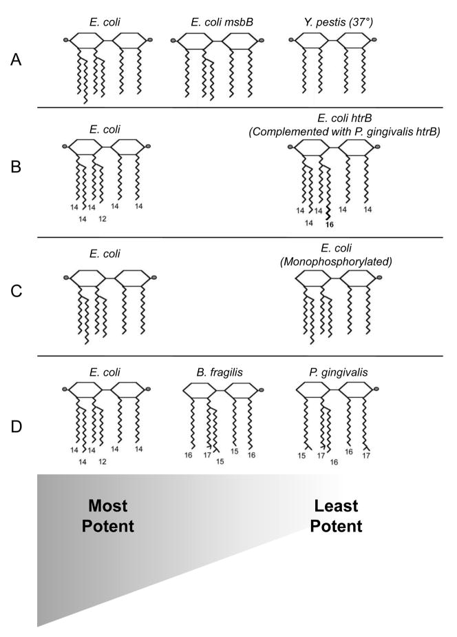
Lipid A structural modifications can substantially impact the host's inflammatory response to bacterial LPS. Bacteroides fragilis, an opportunistic pathogen associated with life-threatening sepsis and intra-abdominal abscess formation, and Bacteroides thetaiotaomicron, a symbiont pivotal for proper host intestinal tissue development, both produce an immunostimulatory LPS comprised of penta-acylated lipid A. Under defined conditions, Porphyromonas gingivalis, an oral pathogen associated with periodontitis, also produces an LPS bearing a penta-acylated lipid A. However, this LPS preparation is 100-1000 times less potent than Bacteroides LPS in stimulating endothelial cells. We analyzed Bacteroides and P. gingivalis lipid A structures using MALDI-TOF MS and gas chromatography to determine the structural basis for this phenomenon. Even though both Bacteroides and P. gingivalis lipid A molecules are penta-acylated and mono-phosphorylated, subtle differences in mass and fatty acid content could account for the observed difference in LPS potency. This fatty acid heterogeneity is also responsible for the peak "clusters" observed in the mass spectra and obfuscates the correlation between LPS structure and immunostimulatory ability. Further, we show the difference in potency between Bacteroides and P. gingivalis LPS is TLR4-dependent. Altogether, the data suggest subtle changes in lipid A structure may profoundly impact the host's innate immune response.


Figures

Fig. 1
Fig. 1
The published structures of (A) E. coli, (B) B. fragilis, and (C) P. gingivalis lipid A. As calculated from their published structures, the masses of each lipid A moiety are 1798, 1662, and 1690 amu, respectively.
Fig. 2
Fig. 2
The MALDI-TOF MS profiles of lipid A isolated from Bacteroides and P. gingivalis. B. fragilis lipid A isolated using (A) the TRI Reagent method or (B) the hot phenol-water method yielded nearly identical MALDI-TOF MS profiles, indicating both methods isolate similar molecules. Comparable results were observed with B. thetaiotaomicron lipid A isolated using (C) the TRI Reagent method or (D) the hot phenol-water method. (E) The MALDI-TOF MS profile of the lipid A isolated from a P. gingivalis mutant capable of only synthesizing a penta-acylated moiety is shown.
Fig. 2
Fig. 2
The MALDI-TOF MS profiles of lipid A isolated from Bacteroides and P. gingivalis. B. fragilis lipid A isolated using (A) the TRI Reagent method or (B) the hot phenol-water method yielded nearly identical MALDI-TOF MS profiles, indicating both methods isolate similar molecules. Comparable results were observed with B. thetaiotaomicron lipid A isolated using (C) the TRI Reagent method or (D) the hot phenol-water method. (E) The MALDI-TOF MS profile of the lipid A isolated from a P. gingivalis mutant capable of only synthesizing a penta-acylated moiety is shown.
Fig. 3
Fig. 3
E-selectin expression profile for LPS-stimulated HUVECS. HUVECs were stimulated with B. fragilis or B. thetaiotaomicron LPS that had been isolated using either the TRI Reagent method or the hot phenol-water method, and E-selectin protein expression was measured by ELISA. Results are means and standard deviations of triplicate wells and are representative of at least two independent determinations.
Fig. 4
Fig. 4
E-selectin expression profile for LPS-stimulated HUVECs. HUVECs were stimulated with E. coli, E. coli msbB, B. fragilis, or B. thetaiotaomicron LPS, and E-selectin protein expression was measured by ELISA. Results are means and standard deviations of triplicate wells and are representative of at least two independent determinations.
Fig. 5
Fig. 5
(A) PVDF membrane stained using colloidal gold. LPS or lipid A samples (10 μg) were run on an SDS-PAGE gel, transferred to a PVDF membrane, and stained with colloidal gold to detect protein contamination. The contents of the lanes were as follows: Protein ladder (lane L); B. thetaiotaomicron LPS (lane 1) or lipid A (lane 2); Pg 1690 LPS (lane 3) or lipid A (lane 4); and 100 ng (lane 5), 10 ng (lane 6), or 1 ng (lane 7) of BSA. (B) E-selectin expression profile for LPS- or lipid A-stimulated HUVECs. HUVECs were stimulated with B. thetaiotaomicron or Pg 1690 LPS or lipid A, and E-selectin protein expression was measured by ELISA. Results are means and standard deviations of triplicate wells and are representative of at least two independent determinations.
Fig. 6
Fig. 6
NF-κB activation profile of TLR4-transfected HEK 293 cells following LPS stimulation. TLR4-transfected HEK 293 cells were stimulated with B. fragilis, B. thetaiotaomicron, or Pg 1690 LPS, and the relative amount of firefly luciferase activity was determined. Values are reported as the fold increase of relative luciferase units (firefly luciferase/Renilla luciferase) for the LPS-stimulated samples compared to the non-stimulated control response, whose ratio was set at 1. Results are means and standard deviations of triplicate wells and are representative of at least two independent determinations.
Fig. 7
Fig. 7
Proposed model for the relationship between lipid A structure and potency. While exceptions to our proposed model certainly exist, the model is meant to reflect general trends in the literature. See Sections 1 and 3 for specific details about each of the lipid A structures. (The lipid A diagrams were graciously provided by Erridge et al.[1]). In each row, the lipid A molecules are arranged from most potent (left) to least potent (right). (A) E. coli, with six fatty acids, is the most potent lipid A structure known. Existing evidence indicates that as the number of attached fatty acids decreases, so does the potency. (B) Lipid A appears to be most potent when the attached fatty acids are 12–14 carbon units long. Deviations from these carbon-chain lengths decrease the potency. (C) Current research indicates the most potent lipid A molecules contain two phosphate groups, and lipid A molecules with one (or no) phosphate groups are much less potent. (D) Data from this work shows that Bacteroides LPS is less potent than E. coli LPS, and P. gingivalis LPS is the least potent of all three. A possible explanation for this is that although both Bacteroides and P. gingivalis possess penta-acylated and mono-phosphorylated lipid A molecules, the fatty acid chain lengths of Bacteroides lipid A are closer in length to those of E. coli lipid A than the fatty acid chain lengths of P. gingivalis lipid A are to those of E. coli lipid A.

References

    1. Erridge C, Bennett-Guerrero E, Poxton IR. Structure and function of lipopolysaccharides. Microbes Infect. 2002;4:837–51. - PubMed
    1. Wilson M. Microbial Inhabitants of Humans. Cambridge, UK: Cambridge University Press; 2005.
    1. Kuwahara T, Yamashita A, Hirakawa H, Nakayama H, Toh H, Okada N, et al. Genomic analysis of Bacteroides fragilis reveals extensive DNA inversions regulating cell surface adaptation. Proc Natl Acad Sci U S A. 2004;101:14919–24. - PMC - PubMed
    1. Fille M, Mango M, Lechner M, Schaumann R. Bacteroides fragilis group: trends in resistance. Curr Microbiol. 2006;52:153–7. - PubMed
    1. Falagas ME, Siakavellas E. Bacteroides, Prevotella, and Porphyromonas species: a review of antibiotic resistance and therapeutic options. Int J Antimicrob Agents. 2000;15:1–9. - PubMed

Publication types

MeSH terms

LinkOut - more resources