

Physiological recordings: basic concepts and implementation during functional magnetic resonance imaging
- PMID: 19460445
- PMCID: PMC2741582
- DOI: 10.1016/j.neuroimage.2009.05.033
Physiological recordings: basic concepts and implementation during functional magnetic resonance imaging
Abstract
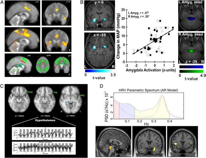
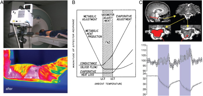
Combining human functional neuroimaging with other forms of psychophysiological measurement, including autonomic monitoring, provides an empirical basis for understanding brain-body interactions. This approach can be applied to characterize unwanted physiological noise, examine the neural control and representation of bodily processes relevant to health and morbidity, and index covert expression of affective and cognitive processes to enhance the interpretation of task-evoked regional brain activity. In recent years, human neuroimaging has been dominated by functional magnetic resonance imaging (fMRI) studies. The spatiotemporal information of fMRI regarding central neural activity is valuably complemented by parallel physiological monitoring, yet such studies still remain in the minority. This review article highlights fMRI studies that employed cardiac, vascular, respiratory, electrodermal, gastrointestinal and pupillary psychophysiological indices to address specific questions regarding interaction between brain and bodily state in the context of experience, cognition, emotion and behaviour. Physiological monitoring within the fMRI environment presents specific technical issues, most importantly related to safety. Mechanical and electrical hazards may present dangers to scanned subjects, operator and/or equipment. Furthermore, physiological monitoring may interfere with the quality of neuroimaging data, or itself be compromised by artefacts induced by the operation of the scanner. We review the sources of these potential problems and the current approaches and advice to enable the combination of fMRI and physiological monitoring in a safe and effective manner.
Figures
References
-
- Abächerli R., Hornaff S., Leber R., Schmid H.J., Felblinger J. Improving automatic analysis of the electrocardiogram acquired during magnetic resonance imaging using magnetic field gradient artefact suppression. J. Electrocardiol. 2006;39:134–139. - PubMed
-
- Abi-Abdallah D., Robin V., Drochon A., Fokapu O. Alterations in human ECG due to the magnetohydrodynamic effect: a method for accurate R peak detection in the presence of high MHD artefacts. Conf. Proc. IEEE Eng. Med. Biol. Soc. 2007:1842–1845. - PubMed
-
- Adair E.R., Black D.R. Thermoregulatory responses to RF energy absorption. Bioelectromagnetics. 2003;6:17–38. - PubMed
-
- Allen P.J., Polizzi G., Krakow K., Fish D.R., Lemieux L. Identification of EEG events in the MR scanner: the problem of pulse artefact and a method for its subtraction. NeuroImage. 1998;8:229–239. - PubMed
-
- Allen P.J., Josephs O., Turner R. A method for removing imaging artefact from continuous EEG recorded during functional MRI. NeuroImage. 2000;12:230–239. - PubMed
Publication types
MeSH terms
Grants and funding
- K01-MH070616/MH/NIMH NIH HHS/United States
- WT_/Wellcome Trust/United Kingdom
- K01 MH070616/MH/NIMH NIH HHS/United States
- R01 HL089850/HL/NHLBI NIH HHS/United States
- K01-AT002166/AT/NCCIH NIH HHS/United States
- P01-AT002048/AT/NCCIH NIH HHS/United States
- K01 AT002166/AT/NCCIH NIH HHS/United States
- P01 AT002048/AT/NCCIH NIH HHS/United States
- F05 AT003770/AT/NCCIH NIH HHS/United States
- R01 AT004714/AT/NCCIH NIH HHS/United States
- R01-HL 089850/HL/NHLBI NIH HHS/United States
- R01-AT004714/AT/NCCIH NIH HHS/United States
- F05-AT003770/AT/NCCIH NIH HHS/United States
LinkOut - more resources
Full Text Sources
Other Literature Sources
Medical
