. 2009 May 22;324(5930):1084-7.
doi: 10.1126/science.1169626.

The human K-complex represents an isolated cortical down-state

Affiliations

The human K-complex represents an isolated cortical down-state

Sydney S Cash et al. Science. .

Abstract

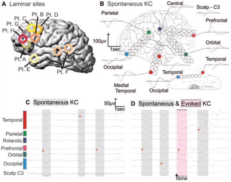
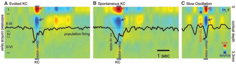
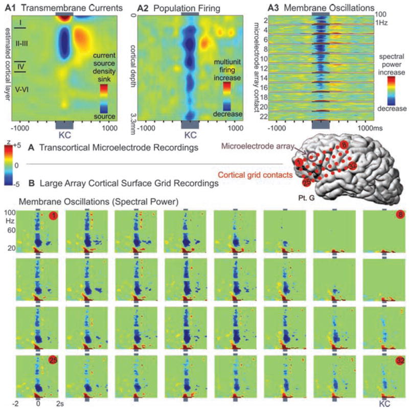
The electroencephalogram (EEG) is a mainstay of clinical neurology and is tightly correlated with brain function, but the specific currents generating human EEG elements remain poorly specified because of a lack of microphysiological recordings. The largest event in healthy human EEGs is the K-complex (KC), which occurs in slow-wave sleep. Here, we show that KCs are generated in widespread cortical areas by outward dendritic currents in the middle and upper cortical layers, accompanied by decreased broadband EEG power and decreased neuronal firing, which demonstrate a steep decline in network activity. Thus, KCs are isolated "down-states," a fundamental cortico-thalamic processing mode already characterized in animals. This correspondence is compatible with proposed contributions of the KC to sleep preservation and memory consolidation.


Figures

Fig. 1
Fig. 1
Spontaneous and evoked KCs recorded simultaneously from the surface of all cortical lobes. (A) Approximate locations of microelectrode arrays in the eight patients (Pt.) studied. (B) Averaged spontaneous KCs recorded with grid electrodes in multiple cortical locations and from the scalp (subject E). Unaveraged spontaneous (C and D) and evoked (D) KCs from the same electrodes. Red asterisks show earliest deflection during the KCs and demonstrate the variability in onset and spread. Sleep stages and KCs were identified during natural sleep with standard criteria (27). KCs were evoked with occasional tones. Subdural electrode arrays were placed to confirm the hypothesized seizure focus and to locate epileptogenic tissue in relation to essential cortex, so as to direct surgical treatment.
Fig. 2
Fig. 2
Population transmembrane currents and neuronal firing in different cortical layers during single evoked (A) and spontaneous (B) KC in stage 2 sleep, and (C) during the slow oscillation in stage 3 to 4 sleep. Evoked and spontaneous KC and the slow-oscillation down-state are all characterized by outward currents (sources) in cortical layers II to III (blue arrows), paired with inward currents (sinks) near the cortical surface (red arrows), and decreased neuronal firing (black arrows). Unlike the KC, the slow-oscillation down-state is embedded in rhythmic alternation with up-states consisting of layer II/III sinks (*), layer I sources (*), and increased firing. Patient F.
Fig. 3
Fig. 3
Decrement in population firing and high-frequency membrane oscillations during KC. (A) Microelectrode array recordings of (A1) averaged spontaneous KCs show a layer III source, (A2) decreased neuronal firing, and (A3) membrane oscillations (higher-frequency spectral power), in all layers, especially upper layers. (B) Simultaneous recordings in the same patient from a large grid array of electrodes on the cortical surface also show a decrease in high-frequency spectral power during KC. Plots are set to the same threshold to show significant (P < 0.01) spectral power changes compared with a period of 1500 to 250 ms before the KC.

Comment in

Similar articles

Cited by

References

    1. Buzsaki G. Rhythms of the Brain. Oxford Univ. Press; Oxford: 2006.
    1. Loomis AL, Harvey EN, Hobart G. J Neurophysiol. 1938;13(suppl):231.
    1. Colrain IM. Sleep. 2005;28:255. - PubMed
    1. Halasz P. Sleep Med Rev. 2005;9:391. - PubMed
    1. Steriade M, Amzica F, Nunez A. J Neurophysiol. 1993;70:1385. - PubMed

Publication types