. 2009 Jun 19;104(12):1355-63.
doi: 10.1161/CIRCRESAHA.108.192880. Epub 2009 May 21.

Lipoprotein accumulation in macrophages via toll-like receptor-4-dependent fluid phase uptake

Affiliations

Lipoprotein accumulation in macrophages via toll-like receptor-4-dependent fluid phase uptake

Soo-Ho Choi et al. Circ Res. .

Abstract

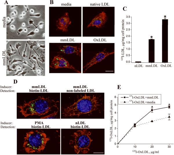
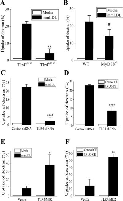
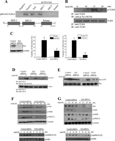
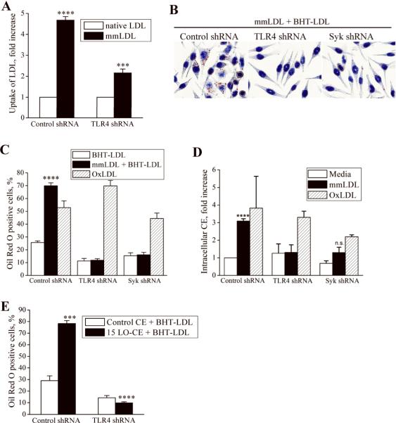
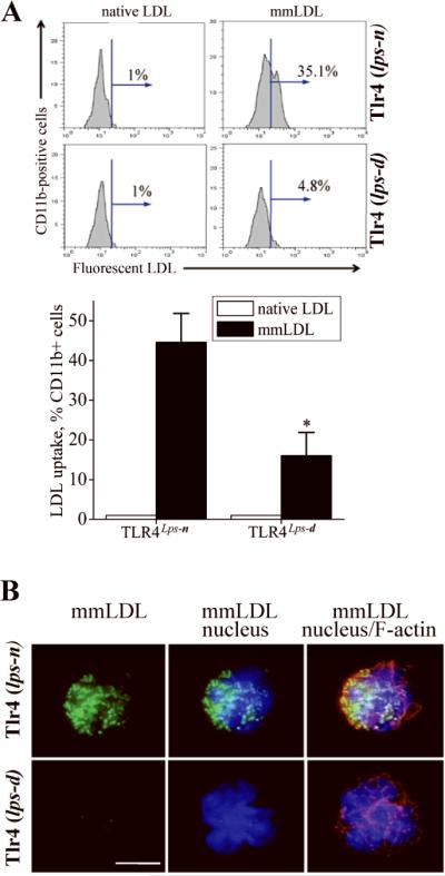
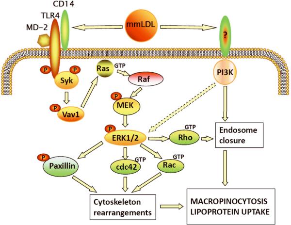
Toll-like receptor (TLR)4 recognizes microbial pathogens, such as lipopolysaccharide, and mediates lipopolysaccharide-induced proinflammatory cytokine secretion, as well as microbial uptake by macrophages. In addition to exogenous pathogens, TLR4 recognizes modified self, such as minimally oxidized low-density lipoprotein (mmLDL). Here we report that mmLDL and its active components, cholesteryl ester hydroperoxides, induce TLR4-dependent fluid phase uptake typical of macropinocytosis. We show that mmLDL induced recruitment of spleen tyrosine kinase (Syk) to a TLR4 signaling complex, TLR4 phosphorylation, activation of a Vav1-Ras-Raf-MEK-ERK1/2 signaling cascade, phosphorylation of paxillin, and activation of Rac, Cdc42, and Rho. These mmLDL-induced and TLR4- and Syk-dependent signaling events and cytoskeletal rearrangements lead to enhanced uptake of small molecules, dextran, and, most importantly, both native and oxidized LDL, resulting in intracellular lipid accumulation. An intravenous injection of fluorescently labeled mmLDL in wild-type mice resulted in its rapid accumulation in circulating monocytes, which was significantly attenuated in TLR4-deficient mice. These data describe a novel mechanism leading to enhanced lipoprotein uptake in macrophages that would contribute to foam cell formation and atherosclerosis. These data also suggest that cholesteryl ester hydroperoxides are an endogenous ligand for TLR4. Because TLR4 is highly expressed on the surface of circulating monocytes in patients with chronic inflammatory conditions, and cholesteryl ester hydroperoxides are present in plasma, lipid uptake by monocytes in circulation may contribute to the pathological roles of monocytes in chronic inflammatory diseases.


Figures

Figure 1
Figure 1. mmLDL-induced uptake of small molecules and native and modified LDL
A, Peritoneal resident macrophages harvested from C57BL6 mice were incubated with media alone or 50 μg/ml mmLDL for 1 hour and imaged with a phase contrast microscope. B, Peritoneal macrophages harvested from C57BL6 mice were incubated with 50 μg/ml of mmLDL, native LDL or OxLDL for 1 hour, in the presence of 50 μg/ml Lucifer Yellow AC. At the end of incubation, the cells were fixed with formaldehyde and stained for Lucifer Yellow (green), nuclei (blue) and F-actin (red). Scale, 10 μm. C, Specific cell degradation of iodinated (125I) native LDL,mmLDL and OxLDL by resident peritoneal macrophages was used as a measure of LDL uptake. *, p<0.01 mmLDL vs. native LDL; 3 independent experiments. D, Uptake of biotinylated native LDL (200 μg/ml) was induced by incubating J774 macrophages for 1 hour with either mmLDL (50 μg/ml) or PMA (1 μg/ml). Biotinylated LDL, green; F-actin cytoskeleton, red; and nuclei, blue. Replacing mmLDL with native LDL, as an inducer of macropinocytosis, resulted in no uptake of biotinylated LDL (lower right panel). Specificity of the biotin-streptavidin staining was controlled by replacing biotinylated LDL with non-labeled LDL (upper right panel). Scale, 10 μm. E, Total degradation (i.e. specific plus non-specific) of 125I-OxLDL (10, 20 and 30 μg/ml) alone in the media or in the presence of 50 μg/ml unlabeled mmLDL. *, p<0.05 (n=3).
Figure 2
Figure 2. TLR4-dependent uptake of dextran
A and B, Peritoneal resident macrophages from TLR4-competent and TLR4-deficient C3H mice (A) and from MyD88-knockout and wild type C57BL6 mice (B) were incubated with media alone or 50 μg/ml mmLDL for 1 hour in the presence of Alexa Fluor-488 labeled dextran (10,000 Da). The dextran uptake was measured by FACS and presented as percent change in the geometric mean of FACS histograms. **, p<0.01 (n=4); #, not significant (p=0.150, n=6). C and D, TLR4-knockdown and control J774 macrophages were incubated with media alone or 50 μg/ml mmLDL for 1 hour (C), or 2.5 μg/ml of non-oxidized CE or 15LO-CE for 15 min (D) in the presence of Alexa Fluor-488 labeled dextran (10,000 Da). ****, p<0.0005 (n=5). E and F, CHO cell lines expressing human TLR4/MD-2 or empty vector were incubated with media alone or 50 μg/ml mmLDL for 1 hour (E), or 2.5 μg/ml of non-oxidized CE or 15LO-CE for 15 min (F) in the presence of Alexa Fluor-488 labeled dextran (10,000 Da). *, p<0.05; **, p<0.01 (n=5).
Figure 3
Figure 3. Signaling of mmLDL-induced, TLR4-dependent macropinocytosis
A, Yeast two-hybrid analysis of binding of the C-terminal domain of TLR4 with the full length Syk, two SH2 domains and the kinase domain of Syk. B, J774 macrophages were incubated with 50 μg/ml mmLDL for indicated times. Cell lysates were immunoprecipitated with a TLR4 antibody and probed with an antibody against phosphotyrosine (upper panel) or TLR4 (lower panel). C, Control and Syk-knockdown J774 macrophages (the Syk-knockdown was confirmed in an immunoblot as shown in left-hand panel) were incubated with media alone or 50 μg/ml mmLDL for 1 hour (middle panel), or 2.5 μg/ml of non-oxidized CE or 15LO-CE for 15 min (right-hand panel), in the presence of Alexa Fluor 488-labeled dextran (10,000 Da). The dextran uptake was measured by FACS and presented as an increase in the geometric mean of FACS histograms compared to media or control CE. Mean±standard error from 3 to 5 independent experiments. *, p<0.05; **, p<0.01. D, Control, Syk-knockdown and TLR4-knockdown J774 macrophages were incubated with 50 μg/ml mmLDL for 30 min. Cell lysates were immunoprecipitated with a Vav1 antibody and probed with an antibody against phospho-tyrosine (upper panel) or Vav1 (lower panel). E, Control, Syk-knockdown and TLR4-knockdown J774 macrophages were incubated with 50 μg/ml mmLDL for 15 min. Cell lysates were tested for Ras-GTP and total Ras. F and G, Control, TLR4-knockdown and Syk-knockdown J774 macrophages were incubated with 50 μg/ml mmLDL for indicated times. Cell lysates were probed for phosphorylated Raf, MEK1 and ERK1/2, and GAPDH as a loading control, as well as for phosphorylated and total paxillin. The Syk knockdown was confirmed by probing the lysates with a Syk antibody, and the TLR4 knockdown was confirmed in a FACS assay as in Online Figure I.
Figure 4
Figure 4. TLR4-dependent uptake of LDL by macrophagesin vitro
A, Control and TLR4-knockdown J774 macrophages were incubated with 50 μg/ml of Alexa Fluor 488 labeled native LDL or mmLDL for 1 hour. The cells were fixed, gently scrapped from the plate and analyzed by FACS for the presence of intracellular LDL. Geometric means of the FACS histograms for mmLDL were normalized to that for native LDL. Mean±standard error from 3 independent experiments. ****, p<0.0001 control/nLDL vs. control/mmLDL; ***, p<0.001 control/mmLDL vs. TLR4 KD/mmLDL. B and C, J774 macrophages, expressing control, TLR4- or Syk-specific shRNA, were incubated for 40 hours with 200 μg/ml native BHT-LDL (LDL protected from oxidation with 10 μM BHT), 50 μg/ml mmLDL plus 200 μg/ml BHT-LDL, or 25 μg/ml OxLDL alone (higher doses of OxLDL cause macrophage apoptosis). At the end of incubation, the cells were stained with Oil Red O. Images in panel B show control, TLR4-knockdown and Syk-knockdown cells incubated with mmLDL+BHT-LDL and correspond to black bars in the graph in panel C. The percentage of cells that displayed positive Oil Red O staining was determined (C). Mean±standard error from 3 independent experiments. ****, p<0.0001 control/mmLDL vs. control/nLDL. D, J774 macrophages, expressing control, TLR4- or Syk-specific shRNA, were incubated for 40 hours with 50 μg/ml mmLDL or 25 μg/ml OxLDL, and intracellular cholesteryl ester (CE) levels were determined. Mean±standard error from 3 independent experiments. ****, p<0.0001 media vs. mmLDL. E, J774 macrophages, expressing control or TLR4-specific shRNA, were incubated for 24 hours with 200 μg/ml native BHT-LDL (LDL protected from oxidation with 10 μM BHT) in the presence of 2.5 μg/ml of control CE or 15LO-CE. At the end of incubation, the cells were stained with Oil Red O. The percentage of cells that displayed positive Oil Red O staining was determined. Mean±standard error from 3 independent experiments. ***, p<0.001 non-oxidized CE vs. 15LO-CE in control cells.
Figure 5
Figure 5. Lipid-loaded circulating monocytes
A, LDLR-/- mice were fed a chow or high fat diet (1.25% cholesterol, 21% milk fat) for 5 weeks. Circulating mononuclear cells were isolated and stained for neutral lipid with Oil Red O. B, Ex vivo monocytes as in A were stained with LipidTox Red to identify neutral lipid (red), Counterstaining with Alexa Fluor488 (actin cytoskeleton, green) and DAPI (nuclei, blue) demonstrates intracellular localization of the lipid. Scale, 5 μm (lipid, F-actin and merge) and 2 μm (merge-zoom). C and D, Numbers of lipid-loaded monocytes as a function of total cholesterol and triglycerides levels in plasma.
Figure 6
Figure 6. TLR4-dependent uptake of LDL by circulating monocytesin vivo
A, Tlr4lps-n and Tlr4lps-d C3H mice were injected with 100 μg of Alexa Fluor 488 labeled native LDL or mmLDL. After 1 hour, blood was collected and the Alexa Fluor 488 fluorescence was detected by FACS in a population of CD11b-positive cells (monocytes). The percentage of Alexa Fluor 488 positive cells was plotted in a bar graph. Mean±standard error from 3 independent experiments. *, p<0.05. B, The monocytes that were analyzed by FACS in panel B, were cytospun on a glass slide and stained with Hoechst 33258 (blue) for nuclei and TRITC-phalloidin (red) for F-actin. The green fluorescence shows intracellular accumulation of Alexa Fluor 488 labeled mmLDL. Scale, 5 μm.
Figure 7
Figure 7. Signaling pathways of mmLDL-induced, TLR4-dependent macropinocytosis and lipoprotein uptake

Similar articles

Cited by

References

    1. Krieger M. The other side of scavenger receptors: pattern recognition for host defense. Curr Opin Lipidol. 1997;8:275–280. - PubMed
    1. Miller YI, Chang MK, Binder CJ, Shaw PX, Witztum JL. Oxidized low density lipoprotein and innate immune receptors. Curr Opin Lipidol. 2003;14:437–445. - PubMed
    1. Michelsen KS, Wong MH, Shah PK, Zhang W, Yano J, Doherty TM, Akira S, Rajavashisth TB, Arditi M. Lack of Toll-like receptor 4 or myeloid differentiation factor 88 reduces atherosclerosis and alters plaque phenotype in mice deficient in apolipoprotein E. PNAS. 2004;101:10679–10684. - PMC - PubMed
    1. Bjorkbacka H, Kunjathoor VV, Moore KJ, Koehn S, Ordija CM, Lee MA, Means T, Halmen K, Luster AD, Golenbock DT, Freeman MW. Reduced atherosclerosis in MyD88-null mice links elevated serum cholesterol levels to activation of innate immunity signaling pathways. Nat Med. 2004;10:416–421. - PubMed
    1. Mullick AE, Tobias PS, Curtiss LK. Modulation of atherosclerosis in mice by Toll-like receptor 2. J Clin Invest. 2005;115:3149–3156. - PMC - PubMed

MeSH terms