

Added value and diagnostic performance of intratumoral susceptibility signals in the differential diagnosis of solitary enhancing brain lesions: preliminary study
- PMID: 19461062
- PMCID: PMC7051626
- DOI: 10.3174/ajnr.A1635
Added value and diagnostic performance of intratumoral susceptibility signals in the differential diagnosis of solitary enhancing brain lesions: preliminary study
Abstract
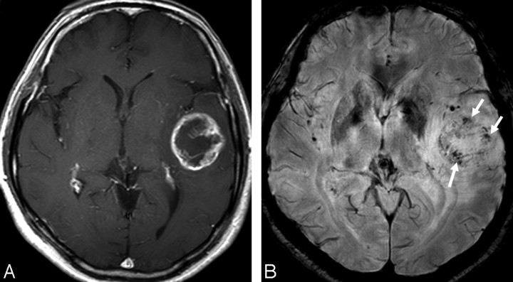
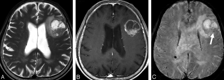
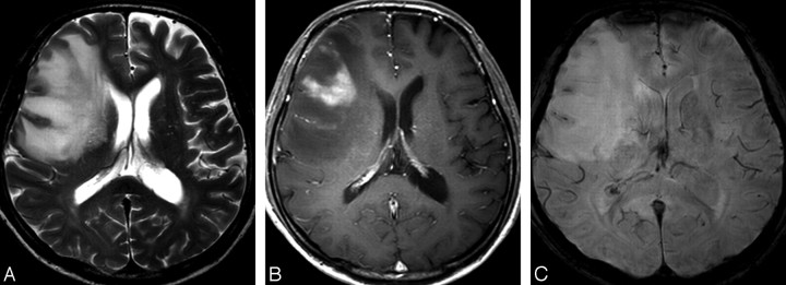
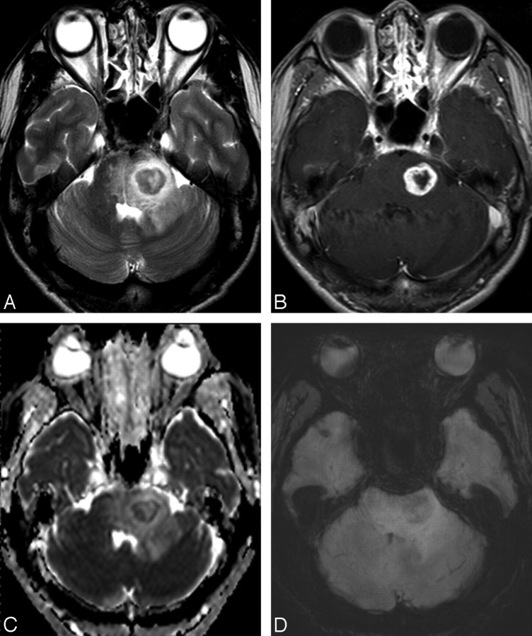
Background and purpose: It has been reported that high-resolution susceptibility-weighted imaging (HR-SWI) is a promising tool for assessing brain tumor characterization noninvasively. The purpose of this study was to determine the added value and diagnostic performance of HR-SWI for differentiating solitary enhancing brain lesions (SELs) by assessing intratumoral susceptibility signals (ITSSs).
Materials and methods: Sixty-four consecutive patients with SELs, without previous surgery, were retrospectively reviewed. We performed 2 consensus reviews, by using conventional MR images alone and with adjunctive HR-SWI. We applied an ITSS grading system based on the degree of the ITSS. Then, we compared the presence and grade of the ITSSs among specific pathologic types of SELs.
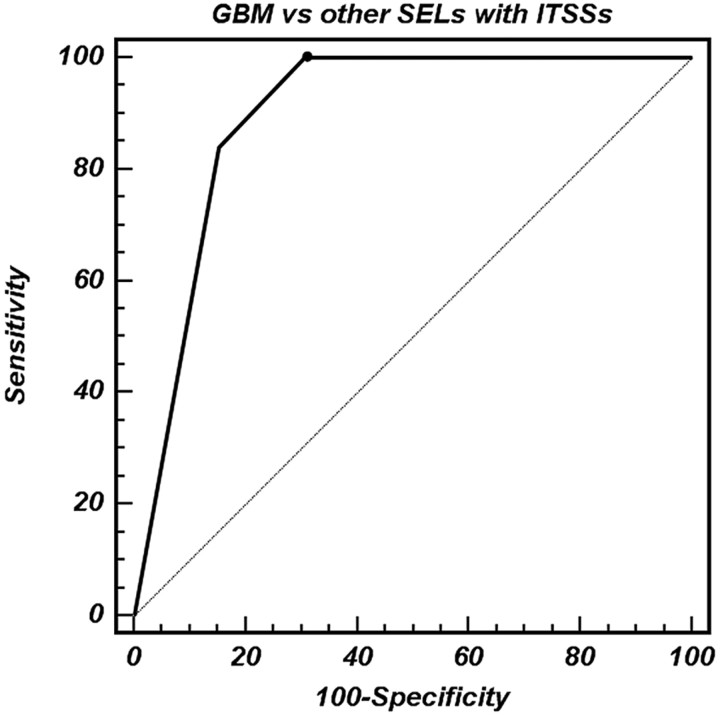
Results: Two observers diagnosed tumor pathology accurately in 43 (67%) of 64 SELs after reviewing the conventional images alone and 50 (78%) of 64 SELs after reviewing the adjunctive HR-SWI (P = .016, McNemar test). ITSSs were seen in 25 (100%) of 25 glioblastoma multiformes (GBMs), in 2 (40%) of 5 anaplastic astrocytomas, and in 11 (73%) of 15 metastatic tumors. Although the ITSSs were unable to distinguish between GBMs and solitary metastatic tumors, differentiation between GBMs and solitary metastatic tumors was achieved (P = .01) by using a high ITSS degree (grade 3). Moreover, the ITSSs could discriminate high-grade gliomas from lymphomas and nontumorous lesions with a specificity of 100% (P < .0001).
Conclusions: The use of ITSSs on HR-SWIs significantly improves the accuracy for the differential diagnosis of SELs compared with the use of conventional MR imaging alone.
Figures

References
-
- Reichenbach JR, Jonetz-Mentzel L, Fitzek C, et al. High-resolution blood oxygen-level dependent MR venography (HRBV): a new technique. Neuroradiology 2001; 43: 364– 69 - PubMed
-
- Reichenbach JR, Essig M, Haacke EM, et al. High-resolution venography of the brain using magnetic resonance imaging. MAGMA 1998; 6: 62– 69 - PubMed
-
- Reichenbach JR, Haacke EM. High-resolution BOLD venographic imaging: a window into brain function. NMR Biomed 2001; 14: 453– 67 - PubMed
-
- Sehgal V, Delproposto Z, Haacke EM, et al. Clinical applications of neuroimaging with susceptibility-weighted imaging. J Magn Reson Imaging 2005; 22: 439– 50 - PubMed
MeSH terms
LinkOut - more resources
Full Text Sources
Medical