

Impact of intra-arterial injection parameters on arterial, capillary, and venous time-concentration curves in a canine model
- PMID: 19461063
- PMCID: PMC7051561
- DOI: 10.3174/ajnr.A1586
Impact of intra-arterial injection parameters on arterial, capillary, and venous time-concentration curves in a canine model
Abstract
Background and purpose: Recent advances in flat panel detector angiographic equipment have provided the opportunity to obtain physiologic and anatomic information from angiographic examinations. To exploit this possibility, one must understand the factors that affect the bolus geometry of an intra-arterial injection of contrast medium. It was our purpose to examine these factors in a canine model.
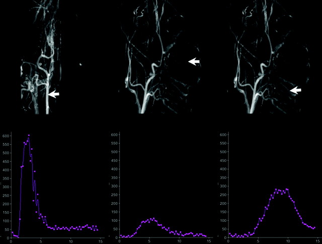
Materials and methods: Under an institutionally approved protocol conforming to Guide for the Care and Use of Laboratory Animals of the National Institutes of Health, 7 canines were placed under general anesthesia with isoflurane and propofol. Through a 5F catheter placed into the right common carotid artery, a series of biplane angiographic acquisitions was obtained to examine the effects caused by variation in the volume of injection, the rate of injection, the duration of injection, the concentration of contrast medium, and the catheter position on arterial, capillary, and venous opacification. The results of each injection protocol were determined from analysis of a time-contrast concentration curve derived from locations over an artery, in brain parenchyma, and over a vein. The curve was generated from 2D digital subtraction angiography acquisitions by using prototype software. The area under the curve, the amplitude of the curve, and the time to peak (TTP) were analyzed separately for each injection parameter.
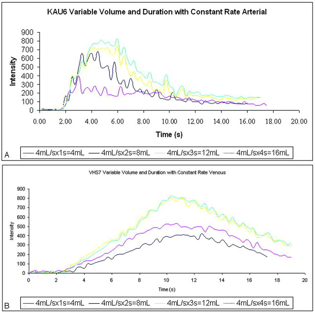
Results: Changes in the injection protocols resulted in predictable changes in the time-concentration curves. The injection parameter that contributed most to maximum opacification was the volume of contrast medium injected. When the injection rate was fixed and the volume was varied, there was an increase in opacification (maximal) proportional to the injected volume. The injected volume also had an indirect (secondary) impact on the temporal characteristics of the opacification. The time-concentration curve became wider, and the peak was shifted to the right as the injection duration increased. The impact of injected volume on maximal opacification was significant (P < .0001), regardless of the site of measurement (artery, tissue, and vein); however, the impact on the temporal characteristics of the time-concentration curve reached statistical significance only in measurements made in the artery and the vein (P < .05), but not in the tissue (P > .1). The impact of injected volume on maximal opacification became nonproportional in the tissue and vein when the volume was very large (>12 mL). Increasing the concentration of contrast medium resulted in a nonproportional increase in the height of the time-concentration curves (P < .05). Injection rate had an impact on both maximal opacification and TTP. The impact on TTP occurred only when the injection rate was very slow (1 mL/s). Changes of concentration had a similar impact on the time-concentration curve. Catheter position did not cause significant alterations in the shape of the curves.
Conclusions: There were predictable effects from modification of injection parameters on the contrast bolus geometry and on time-concentration curves as measured in an artery, brain parenchyma, or a vein. The amplitude, TTP, and area under the time-concentration curve depend mainly and proportionally on the amount of iodine traversing the vasculature per second. Other injection parameters were of less importance in defining bolus geometry. These findings mimic those observed in studies of parameters affecting bolus geometry following an intravenous injection.
Figures
Similar articles
-
Effect of volume and rate of contrast medium injection on intravenous digital subtraction angiographic contrast medium curves.J Am Coll Cardiol. 1984 Aug;4(2):308-15. doi: 10.1016/s0735-1097(84)80219-3. J Am Coll Cardiol. 1984. PMID: 6376594
-
Exploring the Value of Using Color-Coded Quantitative DSA Evaluation on Bilateral Common Carotid Arteries in Predicting the Reliability of Intra-Ascending Aorta Flat Detector CT-CBV Maps.AJNR Am J Neuroradiol. 2015 May;36(5):960-6. doi: 10.3174/ajnr.A4238. Epub 2015 Feb 12. AJNR Am J Neuroradiol. 2015. PMID: 25678483 Free PMC article.
-
Comparison of aortic arch and intravenous contrast injection techniques for C-arm cone beam CT: implications for cerebral perfusion imaging in the angiography suite.Acad Radiol. 2013 Apr;20(4):509-18. doi: 10.1016/j.acra.2012.10.008. Acad Radiol. 2013. PMID: 23498995
-
Optimizing coronary artery opacification and 3D reconstruction from human cadaver hearts in anatomy research.Curr Probl Cardiol. 2024 Feb;49(2):102216. doi: 10.1016/j.cpcardiol.2023.102216. Epub 2023 Nov 20. Curr Probl Cardiol. 2024. PMID: 37993008 Review.
-
Factors Influencing Contrast Enhancement in Abdominal Computed Tomography Angiography in the Dog: A Systematic Review.Animals (Basel). 2024 Dec 5;14(23):3521. doi: 10.3390/ani14233521. Animals (Basel). 2024. PMID: 39682486 Free PMC article. Review.
Cited by
-
Color-coded parametric imaging support display of vessel hemorrhage-an in vitro experiment and clinical validation study.Front Cardiovasc Med. 2024 Jun 20;11:1387421. doi: 10.3389/fcvm.2024.1387421. eCollection 2024. Front Cardiovasc Med. 2024. PMID: 38966753 Free PMC article.
-
Impact of contrast agent injection duration on dynamic contrast-enhanced MRI quantification in prostate cancer.NMR Biomed. 2018 Sep;31(9):e3946. doi: 10.1002/nbm.3946. Epub 2018 Jul 5. NMR Biomed. 2018. PMID: 29974981 Free PMC article.
-
Monitoring of balloon test occlusion of the internal carotid artery by parametric color coding and perfusion imaging within the angio suite: first results.Clin Neuroradiol. 2013 Dec;23(4):285-92. doi: 10.1007/s00062-013-0208-z. Epub 2013 Mar 23. Clin Neuroradiol. 2013. PMID: 23525670 Clinical Trial.
-
Parametric color coding of digital subtraction angiography.AJNR Am J Neuroradiol. 2010 May;31(5):919-24. doi: 10.3174/ajnr.A2020. Epub 2010 Feb 18. AJNR Am J Neuroradiol. 2010. PMID: 20167651 Free PMC article.
-
Relationship between Injection Rate and Contrast Enhancement on Three-dimensional Digital Subtraction Angiography of the Cerebral Arteries.J Belg Soc Radiol. 2018 Dec 3;102(1):76. doi: 10.5334/jbsr.1619. J Belg Soc Radiol. 2018. PMID: 30533599 Free PMC article.
References
-
- Wallace MJ, Kuo MD, Glaiberman C, et al., Three-dimensional C-arm cone-beam CT: applications in the interventional suite. J Vasc Interv Radiol 2008;19:799–813 - PubMed
-
- Burbank FH, Brody WR, Bradley BR. Effect of volume and rate of contrast medium injection on intravenous digital subtraction angiographic contrast medium curves. J Am Coll Cardiol 1984;4:308–15 - PubMed
-
- Cademartiri F, van der Lugt A, Luccichenti G, et al. Parameters affecting bolus geometry in CTA: a review. J Comput Assist Tomogr 2002;26:598–607 - PubMed
MeSH terms
Substances
LinkOut - more resources
Full Text Sources