Review
. 2009 Jun 1;122(Pt 11):1713-21.
doi: 10.1242/jcs.033951.

Molecular mechanisms of clathrin-independent endocytosis

Affiliations
Review

Molecular mechanisms of clathrin-independent endocytosis

Carsten G Hansen et al. J Cell Sci. .

Abstract

There is good evidence that, in addition to the canonical clathrin-associated endocytic machinery, mammalian cells possess multiple sets of proteins that are capable of mediating the formation of endocytic vesicles. The identity, mechanistic properties and function of these clathrin-independent endocytic pathways are currently under investigation. This Commentary briefly recounts how the field of clathrin-independent endocytosis has developed to date. It then highlights recent progress in identifying key proteins that might define alternative types of endocytosis. These proteins include CtBP (also known as BARS), flotillins (also known as reggies) and GRAF1. We argue that a combination of information about pathway-specific proteins and the ultrastructure of endocytic invaginations provides a means of beginning to classify endocytic pathways.


Figures

Fig. 1.
Fig. 1.
Molecular mechanisms for endocytosis. Endocytic pathways can be classified by the ultrastructure of the relevant membrane-transport intermediates as they form at the plasma membrane, coupled with currently emerging information about the molecules that are directly involved in the biogenesis of these intermediates. The electron micrographs shown are our unpublished images (*), or are reproduced from Romer et al. (Romer et al., 2007) (**) and Swanson and Watts (Swanson and Watts, 1995) (***), with permission. Note that the micrograph showing a putative macropinosome is not at the same scale as the others – caveolae are around 60 nm across, the coated pit shown is about 80 nm across, and macropinosomes can be more than 5 μm in diameter.
Fig. 2.
Fig. 2.
Endothelial caveolae in wild-type and caveolin-1–/– mice. Endothelia that line blood vessels [containing red blood cells (RBCs)] were identified in sections of mouse lung studied by transmission electron microscopy. Caveolar structures that are open at the plasma membrane are indicated by arrows in the top panel, and a clathrin-coated pit is also visible in the caveolin-1–/– sample (arrow). These are unpublished data from our laboratory. A more detailed description of endothelial caveolae in caveolin-1–/– mice is given by Zhao et al. (Zhao et al., 2002).
Fig. 3.
Fig. 3.
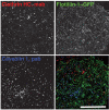
Flotillins, caveolin 1 and clathrin all define different endocytic domains within the plasma membrane. A COS-7 cell expressing flotillin-1–GFP was fixed and stained with a monoclonal antibody (mab) against clathrin heavy chain (HC) and a polyclonal antibody (pab) against caveolin 1. Reproduced from Glebov et al. (Glebov et al., 2006), with permission. Scale bar: 20 nm.

Similar articles

Cited by

References

    1. Aboulaich, N., Vainonen, J. P., Stralfors, P. and Vener, A. V. (2004). Vectorial proteomics reveal targeting, phosphorylation and specific fragmentation of polymerase I and transcript release factor (PTRF) at the surface of caveolae in human adipocytes. Biochem. J. 383, 237-248. - PMC - PubMed
    1. Amstutz, B., Gastaldelli, M., Kalin, S., Imelli, N., Boucke, K., Wandeler, E., Mercer, J., Hemmi, S. and Greber, U. F. (2008). Subversion of CtBP1-controlled macropinocytosis by human adenovirus serotype 3. EMBO J. 27, 956-969. - PMC - PubMed
    1. Barnes, C. J., Vadlamudi, R. K., Mishra, S. K., Jacobson, R. H., Li, F. and Kumar, R. (2003). Functional inactivation of a transcriptional corepressor by a signaling kinase. Nat. Struct. Biol. 10, 622-628. - PubMed
    1. Bashkirov, P. V., Akimov, S. A., Evseev, A. I., Schmid, S. L., Zimmerberg, J. and Frolov, V. A. (2008). GTPase cycle of dynamin is coupled to membrane squeeze and release, leading to spontaneous fission. Cell 135, 1276-1286. - PMC - PubMed
    1. Bast, D. J., Banerjee, L., Clark, C., Read, R. J. and Brunton, J. L. (1999). The identification of three biologically relevant globotriaosyl ceramide receptor binding sites on the Verotoxin 1 B subunit. Mol. Microbiol. 32, 953-960. - PubMed

Publication types