. 2009 Jun 1;122(Pt 11):1834-41.
doi: 10.1242/jcs.045849.

Coatomer-dependent protein delivery to lipid droplets

Affiliations

Coatomer-dependent protein delivery to lipid droplets

Krishnakant G Soni et al. J Cell Sci. .

Abstract

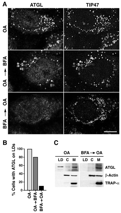
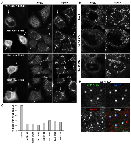
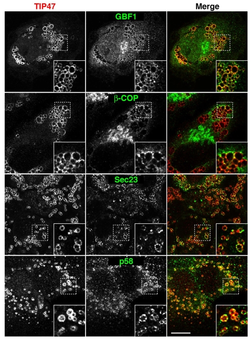
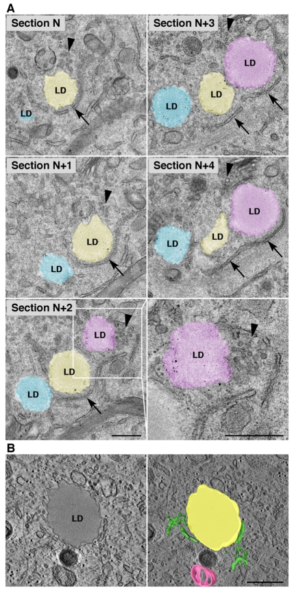
Lipid droplets (LDs) are cytoplasmic organelles that store neutral lipids for use as an energy supply in times of nutrient deprivation and for membrane assembly. Misregulation of LD function leads to many human diseases, including lipodystrophy, obesity and neutral lipid storage disorders. A number of proteins have been shown to localize to the surface of lipid droplets, including lipases such as adipose triglyceride lipase (ATGL) and the PAT-domain proteins ADRP (adipophilin) and TIP47, but the mechanism by which they are targeted to LDs is not known. Here we demonstrate that ATGL and ADRP, but not TIP47, are delivered to LDs by a pathway mediated by the COPI and COPII coatomer proteins and their corresponding regulators.


Figures

Fig. 1.
Fig. 1.
ATGL association with LDs and membranes. (A,B) FRAP analysis of GFP-GGA1 and ATGL-GFP in HeLa cells incubated overnight with 200 μM oleic acid (OA). Boxed regions were bleached at time 0; fluorescence within these regions was monitored at 2-second (GFP-GGA1) or 30-second (ATGL-GFP) intervals. Arrows point to bleached regions. Scale bar: 5 μm. (B) Fluorescence values were plotted as a function of time. Mean and s.d. of at least three independent experiments are shown. (C) HeLa cells were treated with OA as in A, and cell lysates were fractionated into lipid droplets (LD), cytosol (C) and membranes (M) by sucrose gradient centrifugation. Membrane fractions were treated with the indicated agents or left untreated (None). Pellet (P) and supernatant (S) fractions were analyzed by SDS-PAGE and immunoblotting with antibodies to ATGL, TIP47, TRAP-α and calnexin.
Fig. 2.
Fig. 2.
BFA blocks delivery of ATGL to LDs. (A) Cells were treated with 400 μM OA for 3 hours (top row), 400 μM OA for 3 hours followed by 5 μg/ml BFA for 3 hours (middle row), or 5 μg/ml BFA for 10 minutes prior to addition of 400 μM OA and 5 μg/ml BFA for 3 hours (bottom row). Cells were co-immunostained with antibodies to ATGL (left column) and TIP47 (right-hand column). Scale bar: 10 μm. (B) Quantification of results shown in A. At least 100 cells were scored for the presence or absence of ATGL on LDs, and the result plotted as a percentage of total cells examined. (C) Cells were treated with OA in the presence or absence of 1 μg/ml BFA for 19 hours, and lysates were subjected to sucrose gradient fractionation as described in Fig. 1C.
Fig. 3.
Fig. 3.
Depletion of GBF1 inhibits ATGL association with LDs. (A) HeLa cells were transfected with siRNA against GBF1 (knockdown, KD) or mock treated, then co-immunostained with antibodies to ATGL and TIP47. Scale bar: 10 μm. (B) Immunoblot analysis of ATGL levels upon GBF1 KD. HeLa cells were transfected as in A, then treated with 200 μM OA or left untreated, as indicated. Samples were analyzed by SDS-PAGE and immunoblotting with antibodies to ATGL and actin (loading control). Mean and s.d. of at least three independent experiments are shown. (C) Immunoblot analysis of ATGL expression in OA-treated cells incubated in the absence or presence of the proteasomal inhibitor ALLN. The same conditions were used as in B, except that ALLN was added at the same time as OA where indicated. Mean and s.d. of at least three independent experiments are shown. (D) HeLa cells were treated with siRNA against GBF1 in the presence or absence of 400 μM OA. Cells were stained with BODIPY 493/503 and LD diameters measured. Mean and s.d. of at least three independent experiments are shown. (E) Cells were treated as in D and levels of the indicated lipids quantified. Mean and s.d. are shown. In D and E, P-values for significant differences between GBF1-depleted and control cells are indicated. TG, triglyceride; CE, cholesterol ester; FC, free cholesterol; PLs, total phospholipids.
Fig. 4.
Fig. 4.
COPI, COPII and their regulators are required for ATGL delivery to LDs. (A) HeLa cells were transfected with plasmids encoding YFP-GBF1-E794K, ARF1-T31N-GFP, SAR1-T39N-GFP or SAR1-H79G-GFP. At 8 hours after transfection, 150 μM OA was added for an additional 14 hours, followed by co-immunostaining with antibodies to ATGL (middle column) and TIP47 (right-hand column). Arrows indicate cells expressing YFP- or GFP-tagged mutant constructs. (B) HeLa cells were either mock treated (top row) or transfected with siRNAs against β-COP (middle row) or SEC13 (bottom row) and incubated for a total of 72 hours, with 150 μM OA added for the last 12 hours. Cells were immunostained with antibodies to ATGL (left) or TIP47 (right). (C) Quantification of the results shown in Fig. 3A and in A,B. At least 100 cells were scored for the presence or absence of ATGL on LDs, and the result plotted as a percentage of total cells examined. (D) Co-localization of ATGL and ADRP with COPII components at ER exit sites (ERES) in GBF1-KD cells. HeLa cells were treated with GBF1 siRNAs as described in Fig. 2A, transfected with ATGL-GFP, and co-immunostained with antibodies to ADRP and SEC23. Arrows point to SEC23-positive ERES that also contain ATGL and ADRP. Scale bars: 10 μm in A,B; 2 μm in D.
Fig. 5.
Fig. 5.
Immunofluorescence microscopy shows ERES and ER-Golgi intermediate compartment (ERGIC) membranes in close proximity to LDs. HeLa cells were treated for 20 hours with 150 μM OA and co-immunostained with antibodies to TIP47 (red channel, left column) and to GBF1, β-COP, SEC23 or p58 (green channel, middle column). Merged images are shown on the right. Insets are 3-fold magnifications of boxed areas. Scale bar: 10 μm.
Fig. 6.
Fig. 6.
Electron microscopy of ERES and ERGIC structures apposed to LDs. (A) HeLa cells were treated for 20 hours with 150 μM OA and prepared for electron microscopy as described in Materials and Methods. Five serial thin sections through one cell are shown. The bottom right panel is a magnified view of the boxed area in section N+2. Arrows, ER membranes; arrowheads, ERES/ERGIC. (B) Electron tomography showing ERES/ERGC structures in close proximity to a LD. An average of ten tomographic slices, representing a 14 nm virtual section extracted from the tomogram, shows the close proximity of ER and ERES/ERGIC structures to a LD (left). The whole tomogram is 140 nm thick and is shown in supplementary material Movie 1. The volume generated from the tomogram was merged with a single tomogram slice (right). ER and ERES/ERGIC are in green, a LD in yellow, and a mitochondrion in pink. Scale bars: 0.5 μm.
Fig. 7.
Fig. 7.
Model for the involvement of COPII (blue) and COPI (green) in ATGL transport to LDs. ATGL associates with ER membranes and is subsequently transported to specialized ERES and ERGIC structures that are adjacent to pre-existing LDs. ATGL then co-segregates with neutral lipids into nascent LDs, which subsequently fuse with larger LDs. It is also possible that a transfer protein translocates ATGL from an ERGIC domain to LDs. Steps in this process are enabled by sequential action of SAR1-COPII and GBF1-ARF1-COPI.

Similar articles

Cited by

References

    1. Altan-Bonnet, N., Sougrat, R. and Lippincott-Schwartz, J. (2004). Molecular basis for Golgi maintenance and biogenesis. Curr. Opin. Cell Biol. 16, 364-372. - PubMed
    1. Aridor, M., Bannykh, S. I., Rowe, T. and Balch, W. E. (1995). Sequential coupling between COPII and COPI vesicle coats in endoplasmic reticulum to Golgi transport. J. Cell Biol. 131, 875-893. - PMC - PubMed
    1. Arner, P. and Langin, D. (2007). The role of neutral lipases in human adipose tissue lipolysis. Curr. Opin. Lipidol. 18, 246-250. - PubMed
    1. Bannykh, S. I., Rowe, T. and Balch, W. E. (1996). The organization of endoplasmic reticulum export complexes. J. Cell Biol. 135, 19-35. - PMC - PubMed
    1. Bartz, R., Zehmer, J. K., Zhu, M., Chen, Y., Serrero, G., Zhao, Y. and Liu, P. (2007). Dynamic activity of lipid droplets: protein phosphorylation and GTP-mediated protein translocation. J. Proteome Res. 6, 3256-3265. - PubMed

Publication types

MeSH terms

Substances