

Effects of mosapride on motility of the small intestine and caecum in normal horses after jejunocaecostomy
- PMID: 19461212
- PMCID: PMC2801111
- DOI: 10.4142/jvs.2009.10.2.157
Effects of mosapride on motility of the small intestine and caecum in normal horses after jejunocaecostomy
Abstract
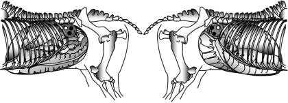
The purpose of the present study was to evaluate the prokinetic effects of mosapride with non-invasive assessment of myoelectrical activity in the small intestine and caecum of healthy horses after jejunocaecostomy. Six horses underwent celiotomy and jejunocaecostomy, and were treated with mosapride (treated group) at 1.5 mg/kg per osos once daily for 5 days after surgery. The other six horses did not receive treatment and were used as controls (non-treated group). The electrointestinography (EIG) maximum amplitude was used to measure intestinal motility. Motility significantly decreased following surgery. In the treated group, the EIG maximum amplitude of the small intestine was significantly higher than in the controls from day 6 approximately 31 after treatment. These findings clearly indicate that mosapride could overcome the decline of intestinal motility after jejunocaecostomy in normal horses.
Figures

Similar articles
-
Effects of mosapride, a 5-hydroxytryptamine 4 receptor agonist, on electrical activity of the small intestine and cecum in horses.Am J Vet Res. 2005 Aug;66(8):1321-3. doi: 10.2460/ajvr.2005.66.1321. Am J Vet Res. 2005. PMID: 16173472
-
Use of multichannel electrointestinography for noninvasive assessment of myoelectrical activity in the cecum and large colon of horses.Am J Vet Res. 2008 Jun;69(6):709-15. doi: 10.2460/ajvr.69.6.709. Am J Vet Res. 2008. PMID: 18518649
-
Effects of mosapride citrate, metoclopramide hydrochloride, lidocaine hydrochloride, and cisapride citrate on equine gastric emptying, small intestinal and caecal motility.Res Vet Sci. 2009 Apr;86(2):302-8. doi: 10.1016/j.rvsc.2008.07.008. Epub 2008 Aug 23. Res Vet Sci. 2009. PMID: 18723200
-
[Pharmacological effects of the gastroprokinetic agent mosapride citrate].Nihon Yakurigaku Zasshi. 1999 May;113(5):299-307. doi: 10.1254/fpj.113.299. Nihon Yakurigaku Zasshi. 1999. PMID: 10480157 Review. Japanese.
-
Gastrointestinal prokinetic benzamides: the pharmacology underlying stimulation of motility.Pharmacol Rev. 1995 Dec;47(4):631-51. Pharmacol Rev. 1995. PMID: 8746557 Review. No abstract available.
Cited by
-
Effects of Single-Dose Prucalopride on Intestinal Hypomotility in Horses: Preliminary Observations.Sci Rep. 2017 Jan 27;7:41526. doi: 10.1038/srep41526. Sci Rep. 2017. PMID: 28128322 Free PMC article.
-
Evaluation of the Effects of Detomidine on Equine Myoelectrical Activity Using Electrointestinography.J Vet Emerg Crit Care (San Antonio). 2025 Mar-Apr;35(2):120-130. doi: 10.1111/vec.13464. Epub 2025 Apr 20. J Vet Emerg Crit Care (San Antonio). 2025. PMID: 40254911 Free PMC article.
References
-
- Bauer AJ, Boeckxstaens GE. Mechanisms of postoperative ileus. Neurogastroenterol Motil. 2004;16(Suppl 2):54–60. - PubMed
-
- Cohen ND, Lester GD, Sanchez LC, Merritt AM, Roussel AJ., Jr Evaluation of risk factors associated with development of postoperative ileus in horses. J Am Vet Med Assoc. 2004;225:1070–1078. - PubMed
-
- Dart AJ, Hodgson DR. Role of prokinetic drugs for treatment of postoperative ileus in the horse. Aust Vet J. 1998;76:25–31. - PubMed
-
- Donawick WJ, Christie BA, Stewart JV. Resection of diseased ileum in the horse. J Am Vet Med Assoc. 1971;159:1146–1149. - PubMed
-
- Embertson RM, Colahan PT, Brown MP, Peyton LC, Schneider RK, Granstedt ME. Ileal impaction in the horse. J Am Vet Med Assoc. 1985;186:570–572. - PubMed
MeSH terms
Substances
LinkOut - more resources
Full Text Sources
