

Defining the cut point between low-grade and high-grade ovarian serous carcinomas: a clinicopathologic and molecular genetic analysis
- PMID: 19461510
- PMCID: PMC2716424
- DOI: 10.1097/PAS.0b013e3181a24354
Defining the cut point between low-grade and high-grade ovarian serous carcinomas: a clinicopathologic and molecular genetic analysis
Abstract
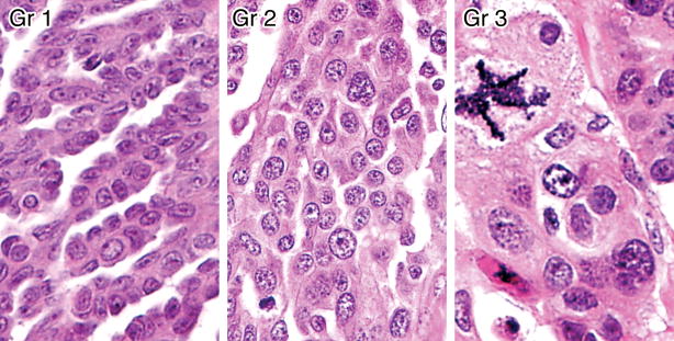
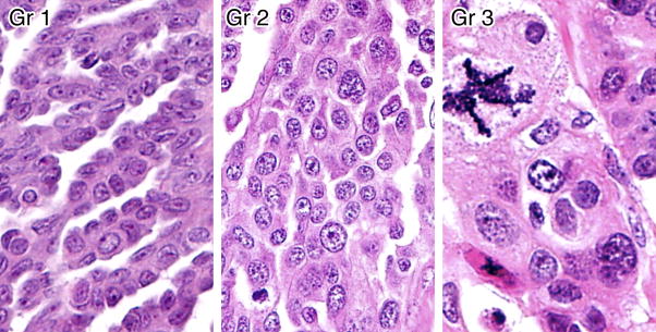
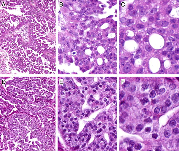
A 2-tier grading system based on nuclear grade divides ovarian serous carcinomas into low (nuclear grade 1) and high grade (nuclear grade 3). In most instances the separation is straightforward but at times, the morphologic distinction between them can be difficult. We studied 11 ovarian serous carcinomas with features that were "intermediate" (nuclear grade 2) between low and high grade. All the cases were high staged and had a poor clinical outcome. None of the tumors showed mutations in KRAS, BRAF, and ERBB2 genes that characterize most low-grade serous carcinomas. In contrast, 10 (90.9%) of 11 cases contained nonsynonymous TP53 mutations characteristic of high-grade serous carcinomas. In summary, the molecular genetic profile and behavior of serous carcinomas with grade 2 nuclei are virtually the same as those of serous carcinomas with grade 3 nuclei, supporting the use of the 2-tier grading system for classifying ovarian serous carcinomas.
Figures

References
-
- Classification and staging of malignant tumours in the female pelvis. Acta Obstet Gynecol Scand. 1971;50:1–7. - PubMed
-
- Campbell IG, Russell SE, Choong DY, et al. Mutation of the PIK3CA gene in ovarian and breast cancer. Cancer Res. 2004;64:7678–81. - PubMed
-
- Gershenson DM, Sun CC, Lu KH, et al. Clinical behavior of stage II-IV low-grade serous carcinoma of the ovary. Obstet Gynecol. 2006;108:361–8. - PubMed
-
- Hsu CY, Kurman RJ, Vang R, et al. Nuclear size distinguishes low- from high-grade ovarian serous carcinoma and predicts outcome. Hum Pathol. 2005;36:1049–54. - PubMed
Publication types
MeSH terms
Substances
Grants and funding
LinkOut - more resources
Full Text Sources
Other Literature Sources
Medical
Research Materials
Miscellaneous
