. 2009 Jun 17;28(12):1803-11.
doi: 10.1038/emboj.2009.138. Epub 2009 May 21.

Structural insight into the essential PB1-PB2 subunit contact of the influenza virus RNA polymerase

Affiliations

Structural insight into the essential PB1-PB2 subunit contact of the influenza virus RNA polymerase

Kanako Sugiyama et al. EMBO J. .

Abstract

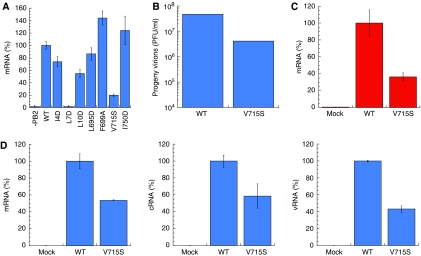
Influenza virus RNA-dependent RNA polymerase is a multi-functional heterotrimer, which uses a 'cap-snatching' mechanism to produce viral mRNA. Host cell mRNA is cleaved to yield a cap-bearing oligonucleotide, which can be extended using viral genomic RNA as a template. The cap-binding and endonuclease activities are only activated once viral genomic RNA is bound. This requires signalling from the RNA-binding PB1 subunit to the cap-binding PB2 subunit, and the interface between these two subunits is essential for the polymerase activity. We have defined this interaction surface by protein crystallography and tested the effects of mutating contact residues on the function of the holo-enzyme. This novel interface is surprisingly small, yet, it has a crucial function in regulating the 250 kDa polymerase complex and is completely conserved among avian and human influenza viruses.


Figures

Figure 1
Figure 1
The nature of the PB1–PB2 interface. (A) Co-precipitation experiments of the C-terminus of PB1 (residues 678–757) co-expressed with different fragments of PB2 carrying a hexa-histidine tag at the N-terminus. The red arrow indicates the PB1-C peptide, which is retained on a Ni-NTA column only with the constructs in lanes 1 (residues 1–37) and 2 (residues 1–86). (B) An overall ribbon diagram showing the structure of the complex, with helices from PB1 coloured red, and helices from PB2 coloured blue. Coil regions are coloured green. (C) The same model as (B), but rotated 90° around a horizontal axis to show the separation between the three helices of the N-terminal peptide of PB2. The principal contacts all involve helix 1 of PB2 (residues 1–12). (D) The sequences of the complexed fragments with a sequence alignment of human (H1N1) influenza, an avian strain (A/Duck/Hong Kong/2000, H5N1), and H7N7 (A/Equine/London/1416/1973). Secondary structure is indicated with red or blue bars showing helices in PB1 and PB2, respectively, and broken lines showing disordered regions. Amino acid residues shown in white on blue form hydrophobic contacts across the PB1–PB2 interface; residues shown in red are not conserved between different viral strains, and, therefore, unlikely to have an essential function. Overall, the interface region between PB1 and PB2 is very highly conserved.
Figure 2
Figure 2
Electron density map. A stereo view of the final electron density map (2mFo-DFc) covering the key residues of the complex. PB1 is shown in red and PB2 in blue. The map has a resolution of 1.7 Å and is contoured at 1.3 σ.
Figure 3
Figure 3
Interactions between PB1 and PB2. (A) A schematic diagram showing the interactions between the two polypeptides. Helix 1 of PB2-N is drawn as a linear model, with the side chains touching PB1 shown in a two-dimensional ball and stick form. Lys 698 and Asp 725 of PB1 form the only salt bridges across the interface, shown as green dotted lines. These salt bridges are not found in every copy of the complex, and mutation studies (replacing Glu 2 or Arg 3 with alanine) show that they have little effect on PB1–PB2 binding (data not shown). Apolar residues of PB1 are shown in red as simple dashed arcs to indicate hydrophobic contacts between 3.4 and3.9 Å in length. This figure was prepared using LIGPLOT (Wallace et al, 1995). (B) A space-filling representation, with PB1 residues shown in yellow and labelled in red. PB2 residues are shown and labelled in blue. The van der Waals surface of each atom is shown semi-transparent. (C) Schematic diagram showing the molecular surface of PB1, coloured by charge (blue positive, red negative). The potential scale ranges from −1 kT/e (red) to 1 kT/e (blue). PB2 is shown as a green ribbon to reveal the PB1-binding surface beneath it is largely apolar. (D) A ribbon diagram showing the helices of PB1-C and PB2-N in red and blue, respectively, with coil regions in green. Side chains selected for mutagenesis are shown as stick models.
Figure 4
Figure 4
RNA synthesis activity of PB1 or PB2 double mutants in HeLa cells. Bar charts showing the level of viral mRNA (A), cRNA (B), or vRNA (C) synthesis by different RNA polymerase double mutants, compared with that of the wild-type (WT) polymerase or with PB2 absent (−PB2). RNA was isolated from HeLa cells transfected with plasmids for expression of viral RNP components. Using primers specific for viral mRNA, cRNA, or vRNA, the production of each RNA type could be separately assessed by quantitative PCR (see Materials and methods). In the absence of the PB2 subunit, enzyme activity is negligible. The results are mean and s.d. for three independent experiments.
Figure 5
Figure 5
RNA synthesis activity of PB1 or PB2 single mutants. (A) Bar chart showing the level of viral mRNA synthesis of different RNA polymerase single mutants compared with that of the wild-type polymerase (WT). The mRNA production in HeLa cells was assayed as in Figure 4A. The L7D mutation effectively abolishes polymerase activity. (B) The yield of progeny virus. MDCK cells were infected with either wild-type or PB1-V715S virus at an MOI=1. After 24 h post infection, the supernatants were collected, and the plaque titer was determined using MDCK cells. The wild-type virus showed roughly 10-fold greater yield than the PB1-V715S mutant. (C) The level of vRNA synthesis in MDCK cells infected with wild-type virus or PB1-V715S virus. MDCK cells were infected with either wild-type or PB1-V715S virus in the presence of 100 μg/ml cycloheximide to block protein synthesis. The real-time quantitative PCR assays were carried out with a primer set specific for NP mRNA, showing that mRNA synthesis is severely curtailed by the single mutation. (D) An identical experiment to (B), but without the addition of cyclohexmide. Production of mRNA (left-hand panel), cRNA (centre) and segment 5 vRNA (right-hand panel) were assayed separately. The mutant polymerase shows significantly reduced activity for each product. The β-actin mRNA was used as an internal control for the whole procedure (see Materials and methods).
Figure 6
Figure 6
Binding assay of PB1 and PB2 mutants. (A) Co-precipitation experiments using PB1-C (residues 678–757) co-expressed with the N-terminus of PB2 (residues 1–86) carrying a hexa-histidine tag at the N-terminus. The complex was loaded on a nickel affinity column and washed before eluting with imidazole and assaying by SDS–PAGE gel. The Coomassie blue stained gel shows that the PB2 fragment is degraded in the absence of PB1 (lane: −PB1). The wild-type PB1 sequence and the V715S mutant both bound strongly to wild-type PB2-N, giving a band for each denatured polypeptide. All the other single mutations tested show significant or complete loss of binding, and consequent degradation of PB2-N. (B) Immunoprecipitation assay of PB1–PB2 interaction. Full-length wild-type PB2, PB2(Δ1–12), and full-length wild-type PB1 were separately expressed in an in vitro (rabbit reticulocyte lysate) translation system (Promega). 35S-methionine labelling was carried out according to the manufacturer's protocol. The recombinant PB1 was incubated with wild-type PB2 (lane 3) or PB2(Δ1–12) (lane 4) at room temperature for 1 h, and then immunoprecipitated using anti-PB2 antibody and protein A-sepharose beads. Protein eluted from the beads was analysed by 7.5% acrylamide SDS–PAGE and visualized by autoradiography. Lane 1 shows PB1 alone, and lane 2 represents a mock experiment. Lane 4 shows a faint band corresponding to PB1, indicating a much weaker interaction with the mutant PB2 than with wild type (lane 3).

References

    1. Barcena J, Ochoa M, de la Luna S, Melero JA, Nieto A, Ortin J, Portela A (1994) Monoclonal antibodies against influenza virus PB2 and NP polypeptides interfere with the initiation step of viral mRNA synthesis in vitro. J Virol 68: 6900–6909 - PMC - PubMed
    1. Biswas SK, Nayak DP (1996) Influenza virus polymerase basic protein 1 interacts with influenza virus polymerase basic protein 2 at multiple sites. J Virol 70: 6716–6722 - PMC - PubMed
    1. Cianci C, Tiley L, Krystal M (1995) Differential activation of the influenza virus polymerase via template RNA binding. J Virol 69: 3995–3999 - PMC - PubMed
    1. Dias A, Bouvier D, Crepin T, McCarthy AA, Hart DJ, Baudin F, Cusack S, Ruigrok RW (2009) The cap-snatching endonuclease of influenza virus polymerase resides in the PA subunit. Nature 458: 914–918 - PubMed
    1. Digard P, Blok VC, Inglis SC (1989) Complex formation between influenza virus polymerase proteins expressed in Xenopus oocytes. Virology 171: 162–169 - PMC - PubMed

Publication types