. 2009 May;5(5):e1000491.
doi: 10.1371/journal.pgen.1000491. Epub 2009 May 22.

Murine leukemias with retroviral insertions at Lmo2 are predictive of the leukemias induced in SCID-X1 patients following retroviral gene therapy

Affiliations

Murine leukemias with retroviral insertions at Lmo2 are predictive of the leukemias induced in SCID-X1 patients following retroviral gene therapy

Utpal P Davé et al. PLoS Genet. 2009 May.

Abstract

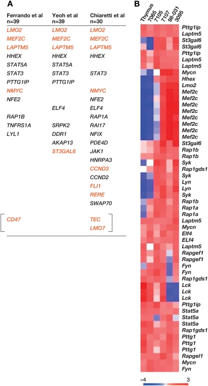
Five X-linked severe combined immunodeficiency patients (SCID-X1) successfully treated with autologous bone marrow stem cells infected ex vivo with an IL2RG-containing retrovirus subsequently developed T-cell leukemia and four contained insertional mutations at LMO2. Genetic evidence also suggests a role for IL2RG in tumor formation, although this remains controversial. Here, we show that the genes and signaling pathways deregulated in murine leukemias with retroviral insertions at Lmo2 are similar to those deregulated in human leukemias with high LMO2 expression and are highly predictive of the leukemias induced in SCID-X1 patients. We also provide additional evidence supporting the notion that IL2RG and LMO2 cooperate in leukemia induction but are not sufficient and require additional cooperating mutations. The highly concordant nature of the genetic events giving rise to mouse and human leukemias with mutations at Lmo2 are an encouraging sign to those wanting to use mice to model human cancer and may help in designing safer methods for retroviral gene therapy.


Conflict of interest statement

The authors have declared that no competing interests exist.

Figures

Figure 1
Figure 1. AKXD Leukemias have frequent clonal Lmo2 insertions of T-cell origin.
A) The mouse Lmo2 gene is shown above the human gene and the five independent viral insertions cloned from AKXD tumors indicated with red arrows. Tumor names are shown above the arrows. The human LMO2 gene has six exons; gray exons are noncoding and yellow are coding. The bracket indicates chromosomal breakpoints in T-ALL involving 11p13. LMO2 was also insertionally activated in four SCID-X gene therapy patients. Only data for patients 4 and 5 from the French trial are available. Red arrows indicate the site and orientation of gene therapy vector insertion. The BamHI sites used for Southern analysis and the positions of probes (A, B, C) are shown. Molecular weight markers are shown to the left of the blots. Retroviral insertions introduce BamHI sites and show a rearranged band indicated with red asterisks. The germline bands were verified by Southern analysis of genomic DNA from brain and indicated G. The blot on the right was done using a probe (p15E) specific to the ecotropic envelope. B) Southern blots using probes specific for immunoglobulin heavy chain Igh and T-cell receptor Jβ1 genes are shown with molecular weight markers. Igh shows a 6 kb germline band and the Jβ1 blot shows a 5.8 kb germline band. The other bands indicate rearrangements that confirm the monoclonality of the tumors. C) Immunohistochemistry of Lmo2-clonal tumors 3095, 7105, and 7107 is shown. The H&E stain shows homogeneous appearing blast cells that have infiltrated and effaced the lymph nodes. All three tumors show mild CD3 staining and mild (3095) to moderate (7105) B220 staining.
Figure 2
Figure 2. Tumor 7107 has two independent insertions in Il2rg and Irs2 genes.
A) LM-PCR identified two independent insertions in tumor 7107, 5′ of the eight coding exons of the Il2rg gene. The two viral chromosomal junctions were independently cloned and sequenced. The insertions were 396 bp apart and should be present in the same clone if they occurred on the same chromosome. Partial sequence for clone 23-10 shows the region of identity with clone 23-1 (green). For clone 23-1, we found viral sequence 5′ of this region instead of chromosome X genomic sequence. B) Two insertions in the Irs2 gene were also isolated from the same tumor. Primers were designed for one allelic insertion for Irs2 and Il2rg and real time PCR performed on the genomic DNA. Primers for a chromosome X sequence were used as reference and show a copy number of 1 for this male mouse. As shown, each insertion comprises about 50% of total tumor genomic DNA. We ruled out polyploidy for Irs2 and Il2rg genes (see Figure S4). C) Panel shows a heat map of gene expression data using the mouse Affymetrix array. The expression of genes near high frequency insertions cloned from 7107 is shown with respect to normal thymus. Log2 intensity scale is shown under the heat map.
Figure 3
Figure 3. Prdm16 and Mef2c genes are frequently insertionally mutated in AKXD Lmo2 tumors.
A) The Prdm16 gene was targeted in tumors 7105 and 7107. Coding exons are in yellow. Tumor 7107 had a clonal insertion as shown by the Southern blot. G shows germline bands and a red asterisk shows the rearrangement. The graph shows quantitative RT-PCR analysis for Prdm16. Real time PCR was normalized to expression in normal thymus. Tumor 7107 had a viral fusion transcript spliced from the 5′LTR to exon 2. The transcript from tumor 7105 was not recovered from the RNA since the insertion was subclonal. B) The Mef2c gene was insertionally activated in three tumors, 3095, 7105, and 98031. Southern blot using BglII-digested genomic DNA using the probe (thick line) shows all insertions were clonal and early in tumorigenesis. Quantitative RT-PCR shows the up-regulation of Mef2c.
Figure 4
Figure 4. Tcf12 insertion creates a truncated protein.
A) Top panel shows the mouse Tcf12 gene with arrows indicating the ubiquitous promoter and an alternate promoter found in thymus (Alt). Yellow exons are coding and the red arrow indicates the retroviral insertion cloned from tumor 3095. B) Panel shows the sequence of the viral fusion transcript cloned by RT-PCR from this tumor. 5′RACE showed transcription was initiated in the viral 5′LTR and spliced into exon 9. The partial sequence of the viral fusion transcript is shown. Red indicates viral sequences, LTR and 5′ of gag. Yellow denotes the start of exon 9 of Tcf12. The first ATG codon (blue) is in frame with the rest of Tcf12. C) Panel shows two SDS-PAGE blots: left panel shows in vitro transcription and translation of the cloned viral fusion cDNA with radiolabeled 35S-methionine. A strong band at 55 kDa appears as well a weaker band at 50 kDa. The in vitro translated proteins were derived from Tcf12 since they were immunoprecipitated with an antibody specific to the protein's COOH-terminus (right panel). D) This heat map shows microarray analysis of the Lmo2-clonal tumors with selected E2A targets. The genes shown are normally up-regulated by E2A except for Gm2a which is repressed. Included in the comparison is tumor 7065 which had a clonal insertion in Notch1 and no up-regulation of Lmo2.
Figure 5
Figure 5. Human microarray studies show consistent high-level expression of genes and pathways insertionally mutated in AKXD Lmo2 tumors.
A) Genes selectively expressed in LMO2-overexpressing patients from three human microarray analyses are shown. Below the first author citations are shown the number of patients in each study. The genes shown in red are retroviral insertions in the mouse Lmo2-clonal tumors. B) Select genes that were expressed in the LMO2-high class of patients were probed in the mouse tumors and are shown in the heat map. Several of these genes (e.g. Mef2c, Hhex, Syk, Lyn, Mycn) are differentially expressed in the mouse tumors in comparison to normal thymus and tumor 7065.

References

    1. Kovanen PE, Leonard WJ. Cytokines and immunodeficiency diseases: critical roles of the gamma(c)-dependent cytokines interleukins 2, 4, 7, 9, 15, and 21, and their signaling pathways. Immunol Rev. 2004;202:67–83. - PubMed
    1. Hacein-Bey-Abina S, Garrigue A, Wang GP, Soulier J, Lim A, et al. Insertional oncogenesis in 4 patients after retrovirus-mediated gene therapy of SCID-X1. J Clin Invest 2008 - PMC - PubMed
    1. Hacein-Bey-Abina S, Von Kalle C, Schmidt M, McCormack MP, Wulffraat N, et al. LMO2-associated clonal T cell proliferation in two patients after gene therapy for SCID-X1. Science. 2003;302:415–419. - PubMed
    1. Deichmann A, Hacein-Bey-Abina S, Schmidt M, Garrigue A, Brugman MH, et al. Vector integration is nonrandom and clustered and influences the fate of lymphopoiesis in SCID-X1 gene therapy. J Clin Invest. 2007;117:2225–2232. - PMC - PubMed
    1. Howe SJ, Mansour MR, Schwarzwaelder K, Bartholomae C, Hubank M, et al. Insertional mutagenesis combined with acquired somatic mutations causes leukemogenesis following gene therapy of SCID-X1 patients. J Clin Invest. 2008;118:3143–3150. - PMC - PubMed

Publication types

MeSH terms