. 2009 May 13:15:962-73.

Ex vivo gene therapy using intravitreal injection of GDNF-secreting mouse embryonic stem cells in a rat model of retinal degeneration

Affiliations

Ex vivo gene therapy using intravitreal injection of GDNF-secreting mouse embryonic stem cells in a rat model of retinal degeneration

Kevin Gregory-Evans et al. Mol Vis. .

Abstract

Purpose: Safe and prolonged drug delivery to the retina is a key obstacle to overcome in the development of new medicines aimed at treating progressive retinal disease. We took advantage of the ability of embryonic stem cells to survive long-term in foreign tissue and used these cells to deliver neuroprotectant molecules to the retina of the rhodopsin TgN S334ter-4 rat model of retinitis pigmentosa (RP).

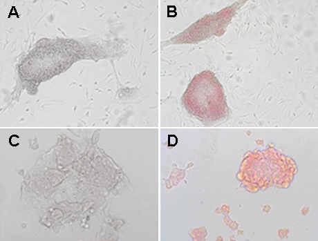
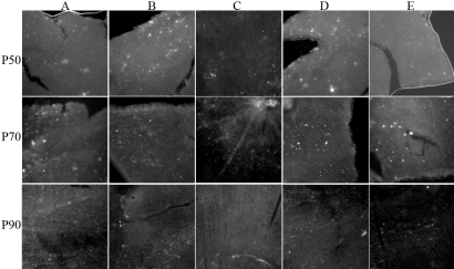
Methods: Mouse embryonic stem (mES) cells, derived from the pluripotent embryonic stem cell line E14TG2a, were genetically engineered to oversecrete the glial cell-derived neurotrophic factor (GDNF). Cell suspensions, containing approximately 200,000 cells and expressing approximately 35ng/10(6) cells/24 h GDNF, were injected into the vitreous cavity of TgN S334ter rat eyes at postnatal day 21 (P21) without immunosuppression. Histological and immunofluorescence imaging was used to evaluate photoreceptor survival up to P90. Local (vitreous) and systemic (serum) concentrations of GDNF were determined and ocular side effects were monitored.

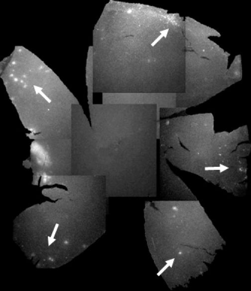
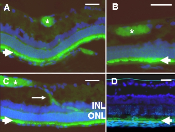
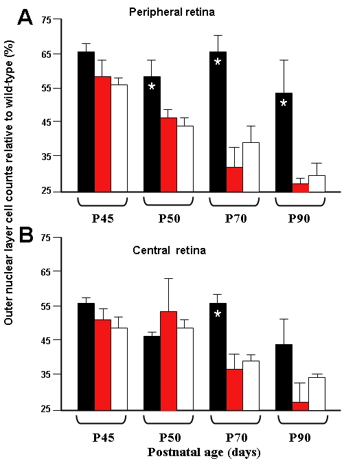
Results: Green fluorescent protein (GFP)-expressing mES cells were observed on the inner limiting membrane of the retina in retinal flatmounts up to P90. In cryostat sections at P45, some GFP-expressing cells had integrated into the inner retina, but did not migrate into the outer nuclear layer. After an initial lag period, the photoreceptor cell counts were significantly higher (p< or =0.05) in animals treated with GDNF-secreting mES cells than in untreated animals, principally in the peripheral retina. Several adverse side effects such as tractional detachments and areas of hyperplasia were seen in a minimal number of treated eyes. Abnormally high levels of GDNF in the peripheral circulation were also observed.

Conclusions: ES cells engineered to secrete GDNF exerted a neuroprotective effect for at least three months on retinal structure in the TgN S334ter rat model of retinal degeneration. Immunosuppression was not required for this. Several adverse effects were identified which require further investigation to make cell-based delivery of neuroprotection a viable clinical strategy.


Figures

Figure 1
Figure 1
GFP and Oct4 expression in mES cells transfected with pGDNF. Cells expressing this GDNF plasmid also express fluorescent protein GFP and will appear red colored when exposed to GFP antibody. A: Control experiment, no red staining if mES cells are not exposed to anti-GFP antibody. B: Immunostaining of mES cells in culture using an anti-GFP antibody gives cells a red coloration confirming GFP cell expression. C: Control experiment, no red staining if mES cells are not exposed to anti- OCT4 antibody. D: Cells immunostained using an anti-human OCT4 (POU5F1) antibody appear red, confirming Oct4 cell expression and establishing cell totipotency.
Figure 2
Figure 2
Composite flatmount images from a single TgN S334ter treated retina at P70. One rat eye from the group treated with intravitreal glial-derived neurotrophic factor (GDNF)-secreting mouse embryonic stem (mES) cells at P21. Spots of increased hyperfluorescence (white), indicative of colonies of green fluorescent protein (GFP)-expressing mES cells, are mainly seen in the peripheral retina (arrows).
Figure 3
Figure 3
Flatmount images of TgN S334ter rat retina treated with GDNF-secreting mES cells. Rows indicate assessments at time points P50, P70, and P90. Columns, represent images from retina from A: upper nasal; B: upper temporal; C: central; D: lower nasal; E: lower temporal areas. All rats were from the group treated with intravitreal glial-derived neurotrophic factor (GDNF)-secreting mouse embryonic stem (mES) cells at P21. White spots indicative of colonies of green fluorescent protein (GFP)-expressing mES cells appear mainly on the surface of the peripheral retina rather than central retina. As time progresses, clumps become more diffuse and ill-defined, suggesting either migration into the retinal tissue or cell loss.
Figure 4
Figure 4
Cryosections of TgN S334ter rat retina studied at post-natal day 35. All sections were immunostained for green fluorescent protein (GFP; green) and retinal cell nuclei were counter-stained blue with DAPI. A-C: Illustrates images from rats from the group treated at post-natal day 21 with intravitreal injection of GDNF-secreting (and GFP-expressing) mouse embryonic stem cells (mES cells). Plates A-C demonstrates integration of GFP-expressing mES cells into the retina. The asterisks mark clumps of mES cells. Some are seen migrating (arrow in C) from the innermost retina toward the outer retina, and extending between the outer nuclear layer (ONL) and inner nuclear layer (INL). D: Illustrates a mock injection eye from the group treated with intravitreal injection of sham injection of PBS, and shows autofluorescence in the retinal pigment epithelium and photoreceptor outer segments (arrowhead). Abbreviations: inner nuclear layer (INL); outer nuclear layer (ONL). Scale bars represent 50 μm.
Figure 5
Figure 5
Treated TgN S334ter rat retina photoreceptor cell nuclei counts. Animals received injections at P21 and cell counts were undertaken at periods up to P90. Graph A represents cell nuclei counts from peripheral retina. Graph B presents cell nuclei counts from posterior pole (central) retina. Cell counts were undertaken in six eyes treated with glial-derived neurotrophic factor (GDNF)-secreting mouse embryonic stem (mES) cells, black bars; in another 8 eyes treated with unmodified mES cells, hatched bars; and in nine eyes treated with sham injections, white bars. Data are presented as the mean percentage of wild-type counts±SEM and analyzed for statistical significance with a Mann–Whitney test. The asterisk indicates p<0.05. The figure demonstrates that preservation of photoreceptor cell nuclei was seen only in eyes treated with GDNF-secreting mES cells and mostly in peripheral retina tissue.

Similar articles

Cited by

References

    1. Travis GH. Mechanisms of cell death in the inherited retinal degenerations. Am J Hum Genet. 1998;62:503–8. - PMC - PubMed
    1. Dunaief JL, Dentchev T, Ying GS, Milam AH. The role of apoptosis in age-related macular degeneration. Arch Ophthalmol. 2002;120:1435–42. - PubMed
    1. Congdon N, O'Colmain B, Klaver CC, Klein R, Muñoz B, Friedman DS, Kempen J, Taylor HR, Mitchell P, Eye Diseases Prevalence Research Group. Causes and prevalence of visual impairment among adults in the United States. Arch Ophthalmol. 2004;122:477–85. - PubMed
    1. Kaur C, Ling EA. Antioxidants and neuroprotection in the adult and developing central nervous system. Curr Med Chem. 2008;15:3068–80. - PubMed
    1. Gonzalez FF, Ferriero DM. Therapeutics for neonatal brain injury. Pharmacol Ther. 2008;120:43–53. - PubMed

Publication types

MeSH terms

Substances