

Genetically encoded fluorescent sensors for studying healthy and diseased nervous systems
- PMID: 19461949
- PMCID: PMC2651031
- DOI: 10.1016/j.ddmod.2008.07.003
Genetically encoded fluorescent sensors for studying healthy and diseased nervous systems
Abstract
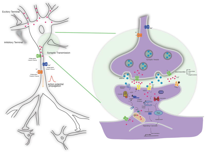
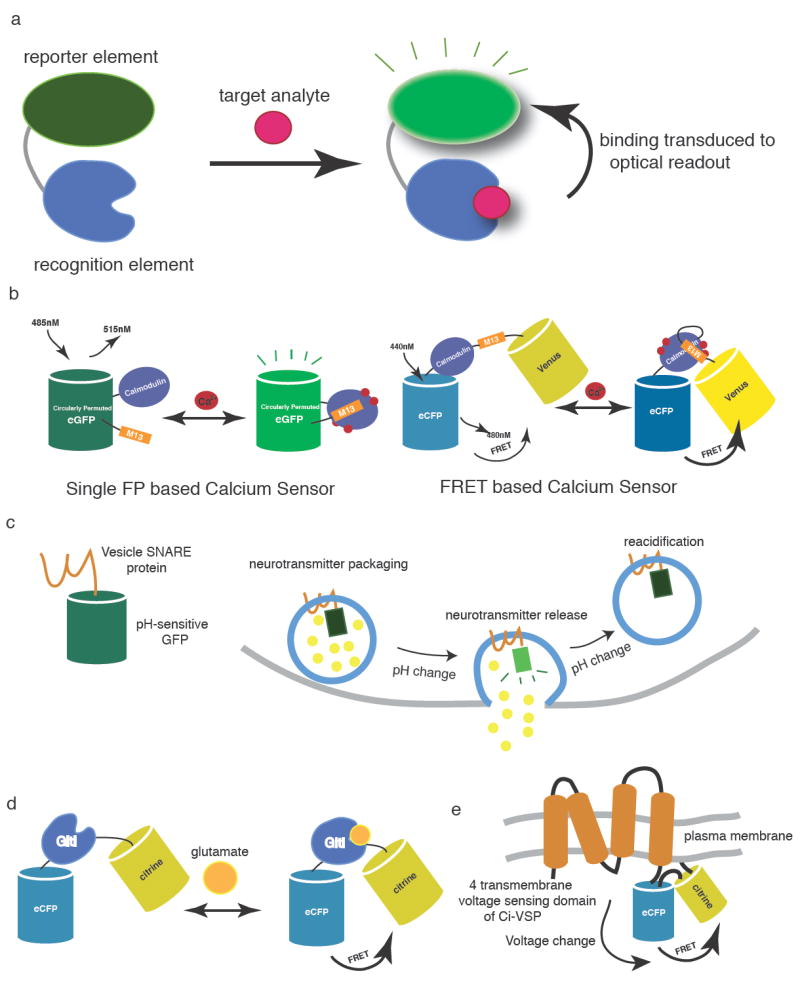
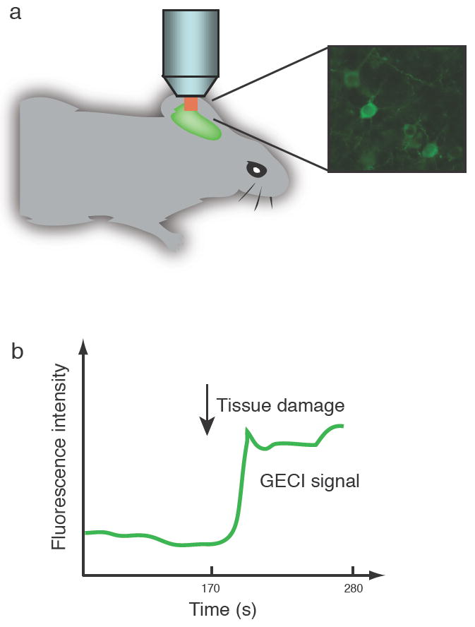
Neurons and glia are functionally organized into circuits and higher-order structures via synaptic connectivity, well-orchestrated molecular signaling, and activity-dependent refinement. Such organization allows the precise information processing required for complex behaviors. Disruption of nervous systems by genetic deficiency or events such as trauma or environmental exposure may produce a diseased state in which certain aspects of inter-neuron signaling are impaired. Optical imaging techniques allow the direct visualization of individual neurons in a circuit environment. Imaging probes specific for given biomolecules may help elucidate their contribution to proper circuit function. Genetically encoded sensors can visualize trafficking of particular molecules in defined neuronal populations, non-invasively in intact brain or reduced preparations. Sensor analysis in healthy and diseased brains may reveal important differences and shed light on the development and progression of nervous system disorders. We review the field of genetically encoded sensors for molecules and cellular events, and their potential applicability to the study of nervous system disease.
Figures
Similar articles
-
Imaging voltage and brain chemistry with genetically encoded sensors and modulators.Curr Opin Chem Biol. 2020 Aug;57:166-176. doi: 10.1016/j.cbpa.2020.07.006. Epub 2020 Aug 18. Curr Opin Chem Biol. 2020. PMID: 32823064 Review.
-
Optical Imaging of Brain Activity In Vivo Using Genetically Encoded Probes.In: Frostig RD, editor. In Vivo Optical Imaging of Brain Function. 2nd edition. Boca Raton (FL): CRC Press/Taylor & Francis; 2009. Chapter 1. In: Frostig RD, editor. In Vivo Optical Imaging of Brain Function. 2nd edition. Boca Raton (FL): CRC Press/Taylor & Francis; 2009. Chapter 1. PMID: 26844329 Free Books & Documents. Review.
-
Engineering Aspects of Olfaction.In: Persaud KC, Marco S, Gutiérrez-Gálvez A, editors. Neuromorphic Olfaction. Boca Raton (FL): CRC Press/Taylor & Francis; 2013. Chapter 1. In: Persaud KC, Marco S, Gutiérrez-Gálvez A, editors. Neuromorphic Olfaction. Boca Raton (FL): CRC Press/Taylor & Francis; 2013. Chapter 1. PMID: 26042329 Free Books & Documents. Review.
-
All-Optical Interrogation of Neural Circuits.J Neurosci. 2015 Oct 14;35(41):13917-26. doi: 10.1523/JNEUROSCI.2916-15.2015. J Neurosci. 2015. PMID: 26468193 Free PMC article.
-
Imaging Voltage in Genetically Defined Neuronal Subpopulations with a Cre Recombinase-Targeted Hybrid Voltage Sensor.J Neurosci. 2017 Sep 20;37(38):9305-9319. doi: 10.1523/JNEUROSCI.1363-17.2017. Epub 2017 Aug 23. J Neurosci. 2017. PMID: 28842412 Free PMC article.
Cited by
-
The single T65S mutation generates brighter cyan fluorescent proteins with increased photostability and pH insensitivity.PLoS One. 2012;7(11):e49149. doi: 10.1371/journal.pone.0049149. Epub 2012 Nov 2. PLoS One. 2012. PMID: 23133673 Free PMC article.
-
Photoactivatable genetically encoded calcium indicators for targeted neuronal imaging.Nat Methods. 2015 Sep;12(9):852-8. doi: 10.1038/nmeth.3480. Epub 2015 Jul 13. Nat Methods. 2015. PMID: 26167640 Free PMC article.
-
Peeking into the sleeping brain: Using in vivo imaging in rodents to understand the relationship between sleep and cognition.J Neurosci Methods. 2019 Mar 15;316:71-82. doi: 10.1016/j.jneumeth.2018.09.011. Epub 2018 Sep 9. J Neurosci Methods. 2019. PMID: 30208306 Free PMC article. Review.
-
Comparison of genetically encoded calcium indicators for monitoring action potentials in mammalian brain by two-photon excitation fluorescence microscopy.Neurophotonics. 2015 Apr;2(2):021014. doi: 10.1117/1.NPh.2.2.021014. Epub 2015 Apr 30. Neurophotonics. 2015. PMID: 26158004 Free PMC article.
-
Live-Cell Imaging of Physiologically Relevant Metal Ions Using Genetically Encoded FRET-Based Probes.Cells. 2019 May 22;8(5):492. doi: 10.3390/cells8050492. Cells. 2019. PMID: 31121936 Free PMC article. Review.
References
Grants and funding
LinkOut - more resources
Full Text Sources