

Tracking and ablating subpopulations of epiblast cells in the chick embryo
- PMID: 19461955
- PMCID: PMC2683548
- DOI: 10.1251/bpo145
Tracking and ablating subpopulations of epiblast cells in the chick embryo
Abstract
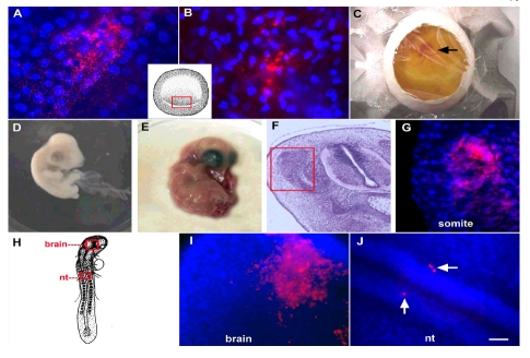
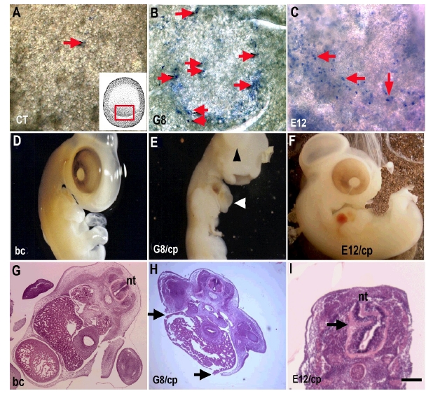
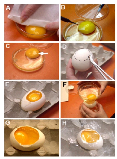
The early chick embryo contains subpopulations of cells that express lineage-specific transcription factors. We have developed protocols to examine the role of these cells during development that involve labeling them for cell tracking purposes and ablating them within the epiblast. The procedures take advantage of the fact that subpopulations of epiblast cells differentially express cell surface antigens recognized by monoclonal antibodies. Embryos are removed from the shell and incubated on the yolk with an antibody. Cells that bind the antibody are either tagged with a fluorescent secondary antibody or lysed with complement. For long-term analyses, embryos are returned to a host shell and placed in an incubator. This method of whole embryo manipulation ex-ovo and incubation in-ovo supports normal development into the fetal period.
Keywords: Chick Embryo; Staining and Labeling.
Figures
Similar articles
-
Chick ex ovo culture and ex ovo CAM assay: how it really works.J Vis Exp. 2009 Nov 30;(33):1620. doi: 10.3791/1620. J Vis Exp. 2009. PMID: 19949373 Free PMC article.
-
Immunohistochemical localisation of monoclonal antibody R 24-recognized ganglioside Glac2 in early chick embryos.Differentiation. 1992 Jan;49(1):7-15. doi: 10.1111/j.1432-0436.1992.tb00764.x. Differentiation. 1992. PMID: 1624061
-
Origin of cells giving rise to mesoderm and endoderm in chick embryo.Nature. 1990 Jan 18;343(6255):273-5. doi: 10.1038/343273a0. Nature. 1990. PMID: 1689008
-
Ex Ovo Culture System for Avian Embryos and its Application.J Poult Sci. 2021 Jan 25;58(1):1-4. doi: 10.2141/jpsa.0200016. J Poult Sci. 2021. PMID: 33519280 Free PMC article. Review.
-
The method of chicken whole embryo culture using the eggshell windowing, surrogate eggshell and ex ovo culture system.Br Poult Sci. 2018 Apr;59(2):240-244. doi: 10.1080/00071668.2017.1413234. Epub 2018 Jan 11. Br Poult Sci. 2018. PMID: 29206486 Review.
Cited by
-
Myo/Nog cell regulation of bone morphogenetic protein signaling in the blastocyst is essential for normal morphogenesis and striated muscle lineage specification.Dev Biol. 2011 Nov 1;359(1):12-25. doi: 10.1016/j.ydbio.2011.08.007. Epub 2011 Aug 18. Dev Biol. 2011. PMID: 21884693 Free PMC article.
-
Noggin producing, MyoD-positive cells are crucial for eye development.Dev Biol. 2009 Dec 1;336(1):30-41. doi: 10.1016/j.ydbio.2009.09.022. Epub 2009 Sep 22. Dev Biol. 2009. PMID: 19778533 Free PMC article.
References
-
- George-Weinstein M, Gerhart J, Gerhart R, Flynn J, Callihan B, Mattiacci M, Miehle C, Foti G, Lash JW, Weintraub W. Skeletal myogenesis: The preferred pathway of chick embryo epiblast cells in vitro. Dev Biol. 1996;173:279–291. - PubMed
-
- Strony R, Gerhart J, Tornambe D, Perlman J, Neely C, Dare J, Stewart B, George- Weinstein M. NeuroM and MyoD are expressed in separate subpopulations of cells in the pregastrulating epiblast. Gene Exp Patterns. 2005;5:387–395. - PubMed
Grants and funding
LinkOut - more resources
Full Text Sources
Other Literature Sources
