. 2009 May 21;4(5):e5651.
doi: 10.1371/journal.pone.0005651.

A new family of receptor tyrosine kinases with a venus flytrap binding domain in insects and other invertebrates activated by aminoacids

Affiliations

A new family of receptor tyrosine kinases with a venus flytrap binding domain in insects and other invertebrates activated by aminoacids

Arnaud Ahier et al. PLoS One. .

Abstract

Background: Tyrosine kinase receptors (RTKs) comprise a large family of membrane receptors that regulate various cellular processes in cell biology of diverse organisms. We previously described an atypical RTK in the platyhelminth parasite Schistosoma mansoni, composed of an extracellular Venus flytrap module (VFT) linked through a single transmembrane domain to an intracellular tyrosine kinase domain similar to that of the insulin receptor.

Methods and findings: Here we show that this receptor is a member of a new family of RTKs found in invertebrates, and particularly in insects. Sixteen new members of this family, named Venus Kinase Receptor (VKR), were identified in many insects. Structural and phylogenetic studies performed on VFT and TK domains showed that VKR sequences formed monophyletic groups, the VFT group being close to that of GABA(B) receptors and the TK one being close to that of insulin receptors. We show that a recombinant VKR is able to autophosphorylate on tyrosine residues, and report that it can be activated by L-arginine. This is in agreement with the high degree of conservation of the alpha amino acid binding residues found in many amino acid binding VFTs. The presence of high levels of vkr transcripts in larval forms and in female gonads indicates a putative function of VKR in reproduction and/or development.

Conclusion: The identification of RTKs specific for parasites and insect vectors raises new perspectives for the control of human parasitic and infectious diseases.


Conflict of interest statement

Competing Interests: The authors have declared that no competing interests exist.

Figures

Figure 1
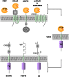
Figure 1. VKR are original proteins composed of a VFTM associated to a TK domain.
The top panel represents the VFTM found in the bacterian periplasmic binding proteins (PBP) and in eukaryotic cell surface membrane receptors (iGluR: ionotropic Glutamate Receptor; ANFR: Atrial Natriuretic Factor Receptor; mGluR: metabotropic Glutamate Receptor; GABABR: metabotropic α-aminobutyric acid Receptor). The bottom panel represents various RTK (EGFR: Epidermal Growth Factor Receptor; FGFR: Fibroblast Growth Factor Receptor; IR: Insulin Receptor composed of α and β chains). VFTM: Venus Flytrap Module, GC, Guanylate Cyclase domain; TK, Tyrosine Kinase domain; CRD: Cystein Rich Domain; IgD: Immunoglobulin Domain; FNIII: FibroNectin III Domain.
Figure 2
Figure 2. Genomic structure of vkr genes.
Genes were ordered following the FlyBase hierarchical tree that showed insect species for which data were available. Predicted exons were represented by rectangles drawn to scale for each vkr gene. Genes of seven Drosophila species (D. ananasse, D. pseudoobscura, D. persimilis, D. wilistoni, D. mojavensis, D. virilis, and D. grimshawi), three mosquitoes (C. pipiens, A. aegypti, A. gambiae), the coleopteran T. castaneum, two hymenopteran species (A. mellifera and N. vitripennis), the phthirapteran P. humanus corporis, the trematode S. mansoni and the echinoderm S. purpuratus are presented. Numbers indicate respective gene lengths in kilobases. Not found indicates that the presence of the vkr gene was not found in the species studied.
Figure 3
Figure 3. Conserved structure of receptors composing the VKR family.
VFTM, TM and TK domains are respectively shown in orange, green and purple. Arrows give the positions of conserved introns in the different sequences with their corresponding numbers in each sequence. The different colors used for arrows indicated variable degrees of conservation for intron positions, as mentioned. The positions of residues delimiting the different domains are indicated by numbers. Red asterisks correspond to VKR for which cDNAs were cloned and sequenced.
Figure 4
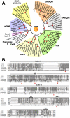
Figure 4. Phylogenetic analyses and sequence alignment of VFTM from VKR.
A-Phylogenetic relationship of VFT protein domains from VKR and VFT-containing receptors, GABAB receptor (GABABR1 and GABABR2 subunits), the natriuretic peptide receptor (ANFR), the metabotropic glutamate-like receptors (mGluR, sweet taste, pheromone and CaSR calcium receptors) and the iGluR N-methyl-D-aspartate (NMDA) receptor mostly from insects and invertebrates. Bootstrap values (10 000 replicates) higher than 80% were shown by a red point on the major internal node only. Genbank accession numbers of receptors used are in table S1. B- Sequence alignment of VFT protein domains from different insect VKRs (AmVKR, TcVKR, AgVKR and DpseuVKR for which cDNA was cloned and sequenced) with the extracellular domain of the human GABABR1 (Q9UBS5) and of the Rattus Norvegicus (NP_058708.1) mGluR5 subunit using the CLUSTAL W method. Residues highlighted in black are those identical in at least 50% of the sequences. Those in grey background correspond to residues homologous in at least 50% of the sequences. The positions highlighted in red with an asterisk are those important for glutamate binding in the mGluR5 subunit.
Figure 5
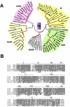
Figure 5. Phylogenetic analyses and sequence alignment of TK domains from VKR.
A-Phylogenetic relationships of TK from VKR and various RTK, the receptors for insulin (IR), the epidermal growth factor (EGFR), the fibroblast growth factor (FGFR) and the proto-oncogene c-ros receptor (ROSR). Bootstrap values (10 000 replicates) higher than 80% were shown by a red point on the major internal node only. Genbank accession numbers of receptors used are in table S1. B- Sequence alignment of the catalytic domains from insect VKR (AmVKR, TcVKR, AgVKR and DpseuVKR) with the TK domain of human insulin receptor (NP_000199) using the CLUSTAL W method. Shaded areas represent residues which are identical (in black) or similar (in grey) in at least 50% of the aligned sequences. Numbers I to XI indicate the eleven subdomains conserved in kinase domains. Consensus sequences required for TK activity are in bold italics.
Figure 6
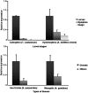
Figure 6. VKR are expressed in larvae and in gonads of adult insects and invertebrates Vkr transcripts were quantified in the different developmental stages of T. castaneum and of A. mellifera drone (A) and in tissues of A. gambiae and S. purpuratus (B).
For graphical representation of qPCR data, cycle thresholds (Δ Ct values) obtained for the different samples were deducted from the Δ Ct value obtained for larval stage (A) or gonad (B) transcript levels. Values were normalized as relative fold-difference using the Δ-Δ Ct (ΔΔ Ct) method. Data are means±s.e. of triplicates from a typical experiment.
Figure 7
Figure 7. AmVKR shows tyrosine kinase activity.
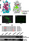
A- Evolutionary conservation of residues of the VKR TK domain (left panel) visualized on the human IR TK crystal structure (PDB accession number 1IR3). Conservation scores are according to a color scale from variable (blue) to conserved (purple) residues. For comparison, the crucial residues needed for kinase activity (right panel) are indicated. B- Expression of the HA-tagged AmVKR in HEK-293 transfected cells revealed by anti-HA antibodies. C- Tyrosine kinase activity of HA-tagged AmVKR proteins. Lysates from HEK293 cells transfected by plasmids containing AmVKR or mutated versions of AmVKR, or by empty prk5 plasmid as a control, were immunoprecipitated by anti-HA antibodies. Kinase assays were performed as described in Materials and Methods. Proteins were analysed by Western blot and tyrosine phosphorylation was detected using P-Tyr-100 antibodies.
Figure 8
Figure 8. VKR can form dimers.
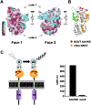
A- Evolutionary conservation of residues at the surface of all VKR VFTMs visualized on both faces of the VFTs (Face 1 and Face 2) using the tridimensional model of AmVKR. Conservation scores are according to a color scale from variable (blue) to conserved (purple) residues. B- Ribbon view of the AmVKR VFTM is shown, with the putative N-glycosylation sites (C-α of Asn residue) found in AmVKR (in red) and in all described VKRs (in orange). C- Time-resolved FRET signal measured between snap-tag (ST) labeled AmVKR subunits at cell surface.
Figure 9
Figure 9. VKR VFTM can bind L-aminoacids responsible for activation.
A- Detailed view of glutamate binding in the VFTM of mGlu1 structure. The five residues (labeled in green) responsible for the binding of the α-amino acid functions of the glutamate, and the equivalent residues in VKRs (in black) are shown. The conserved Lys409 in mGlu1 that interact with the carboxylate function of the glutamate side-chain is displayed, and the corresponding tyrosine residue in VKR is indicated. B- Modulation of recombinant AmVKR protein tyrosine kinase activity by aminoacid ligands. Kinase assays were performed on HA-tagged AmVKR immunoprecipitated from transfected HEK293 cells as described in Fig. 7C. L-Arg or L-Lys (100 µM final concentration) amino-acids were added to AmVKR kinase assay reactions, and the constitutively active AmVKRYYEE proteins were used as positive control.

Similar articles

Cited by

References

    1. Yarden Y, Ullrich A. Growth factor receptor tyrosine kinases. Annu Rev Biochem. 1988;57:443–478. - PubMed
    1. Blume-Jensen P, Hunter T. Oncogenic kinase signalling. Nature. 2001;411:355–365. - PubMed
    1. Plowman GD, Sudarsanam S, Bingham J, Whyte D, Hunter T. The protein kinases of Caenorhabditis elegans: a model for signal transduction in multicellular organisms. Proc Natl Acad Sci U S A. 1999;96:13603–13610. - PMC - PubMed
    1. Miller MA, Steele RE. Lemon encodes an unusual receptor protein-tyrosine kinase expressed during gametogenesis in Hydra. Dev Biol. 2000;224:286–298. - PubMed
    1. Reidling JC, Miller MA, Steele RE. Sweet Tooth, a novel receptor protein-tyrosine kinase with C-type lectin-like extracellular domains. J Biol Chem. 2000;275:10323–10330. - PubMed

Publication types

MeSH terms

Substances