

An active role of the DeltaN isoform of p63 in regulating basal keratin genes K5 and K14 and directing epidermal cell fate
- PMID: 19461998
- PMCID: PMC2680039
- DOI: 10.1371/journal.pone.0005623
An active role of the DeltaN isoform of p63 in regulating basal keratin genes K5 and K14 and directing epidermal cell fate
Abstract
Background: One major defining characteristic of the basal keratinocytes of the stratified epithelium is the expression of the keratin genes K5 and K14. The temporal and spatial expression of these two genes is usually tightly and coordinately regulated at the transcriptional level. This ensures the obligate pairing of K5 and K14 proteins to generate an intermediate filament (IF) network that is essential for the structure and function of the proliferative keratinocytes. Our previous studies have shown that the basal-keratinocyte restricted transcription factor p63 is a direct regulator of K14 gene.
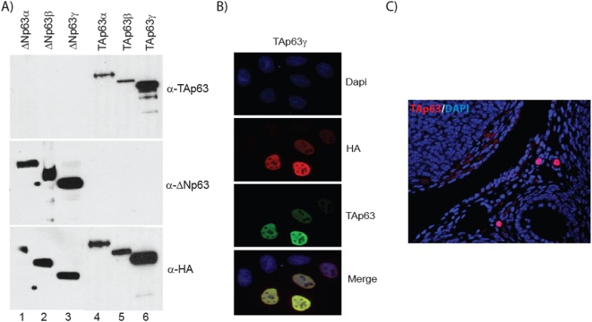
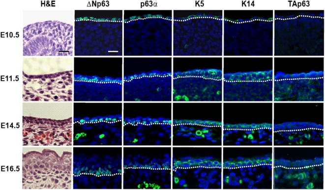
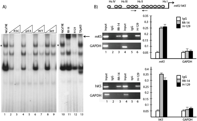
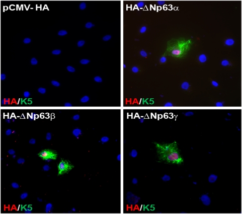
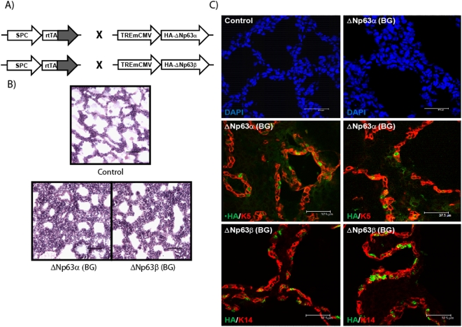
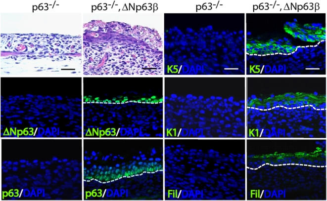
Methodology/principal findings: Here we provide evidence that p63, specifically the DeltaN isoform also regulates the expression of the K5 gene by binding to a conserved enhancer within the 5' upstream region. By using specific antibodies against DeltaNp63, we show a concordance in the expression between basal keratins and DeltaNp63 proteins but not the TAp63 isoforms during early embryonic skin development. We demonstrate, that contrary to a previous report, transgenic mice expressing DeltaNp63 in lung epithelium exhibit squamous metaplasia with de novo induction of K5 and K14 as well as transdifferentiation to the epidermal cell lineage. Interestingly, the in vivo epidermal inductive properties of DeltaNp63 do not require the C-terminal SAM domain. Finally, we show that DeltaNp63 alone can restore the expression of the basal keratins and reinitiate the failed epidermal differentiation program in the skin of p63 null animals.
Significance: DeltaNp63 is a critical mediator of keratinocyte stratification program and directly regulates the basal keratin genes.
Conflict of interest statement
Figures
Similar articles
-
Differential roles of p63 isoforms in epidermal development: selective genetic complementation in p63 null mice.Cell Death Differ. 2006 Jun;13(6):1037-47. doi: 10.1038/sj.cdd.4401926. Cell Death Differ. 2006. PMID: 16601749
-
ΔNp63 knockout mice reveal its indispensable role as a master regulator of epithelial development and differentiation.Development. 2012 Feb;139(4):772-82. doi: 10.1242/dev.071191. Development. 2012. PMID: 22274697 Free PMC article.
-
A functional enhancer of keratin14 is a direct transcriptional target of deltaNp63.J Invest Dermatol. 2007 May;127(5):1175-86. doi: 10.1038/sj.jid.5700652. Epub 2006 Dec 7. J Invest Dermatol. 2007. PMID: 17159913
-
Dynamic life of a skin keratinocyte: an intimate tryst with the master regulator p63.Indian J Exp Biol. 2011 Oct;49(10):721-31. Indian J Exp Biol. 2011. PMID: 22013738 Review.
-
TAp63 and DeltaNp63 in cancer and epidermal development.Cell Cycle. 2007 Feb 1;6(3):274-85. doi: 10.4161/cc.6.3.3797. Epub 2007 Feb 3. Cell Cycle. 2007. PMID: 17264681 Review.
Cited by
-
The WNT-controlled transcriptional regulator LBH is required for mammary stem cell expansion and maintenance of the basal lineage.Development. 2015 Mar 1;142(5):893-904. doi: 10.1242/dev.110403. Epub 2015 Feb 5. Development. 2015. PMID: 25655704 Free PMC article.
-
Comparative transcriptional profiling of the limbal epithelial crypt demonstrates its putative stem cell niche characteristics.BMC Genomics. 2010 Sep 29;11:526. doi: 10.1186/1471-2164-11-526. BMC Genomics. 2010. PMID: 20920242 Free PMC article.
-
Pterygial body epithelium domination of pterygial proliferation with TCF4 as a potential key factor.Int J Ophthalmol. 2018 Sep 18;11(9):1467-1474. doi: 10.18240/ijo.2018.09.07. eCollection 2018. Int J Ophthalmol. 2018. PMID: 30225220 Free PMC article.
-
A Maverick Review of Common Stem/Progenitor Markers in Lung Development.Stem Cell Rev Rep. 2022 Dec;18(8):2629-2645. doi: 10.1007/s12015-022-10422-z. Epub 2022 Jul 23. Stem Cell Rev Rep. 2022. PMID: 35871209 Review.
-
Dysregulated ΔNp63α inhibits expression of Ink4a/arf, blocks senescence, and promotes malignant conversion of keratinocytes.PLoS One. 2011;6(7):e21877. doi: 10.1371/journal.pone.0021877. Epub 2011 Jul 15. PLoS One. 2011. PMID: 21789189 Free PMC article.
References
-
- Coulombe PA, Omary MB. ‘Hard’ and ‘soft’ principles defining the structure, function and regulation of keratin intermediate filaments. Curr Opin Cell Biol. 2002;14:110–122. - PubMed
-
- Koster MI, Roop DR. Mechanisms regulating epithelial stratification. Annu Rev Cell Dev Biol. 2007;23:93–113. - PubMed
-
- Nagarajan P, Romano RA, Sinha S. Transcriptional control of the differentiation program of interfollicular epidermal keratinocytes. Crit Rev Eukaryot Gene Expr. 2008;18:57–79. - PubMed
Publication types
MeSH terms
Substances
Grants and funding
LinkOut - more resources
Full Text Sources
Other Literature Sources
Molecular Biology Databases
Research Materials
Miscellaneous
