. 2009 May 20;4(5):e5622.
doi: 10.1371/journal.pone.0005622.

Polycomb mediated epigenetic silencing and replication timing at the INK4a/ARF locus during senescence

Affiliations

Polycomb mediated epigenetic silencing and replication timing at the INK4a/ARF locus during senescence

Hanane Agherbi et al. PLoS One. .

Abstract

Background: The INK4/ARF locus encodes three tumor suppressor genes (p15(Ink4b), Arf and p16(Ink4a)) and is frequently inactivated in a large number of human cancers. Mechanisms regulating INK4/ARF expression are not fully characterized.

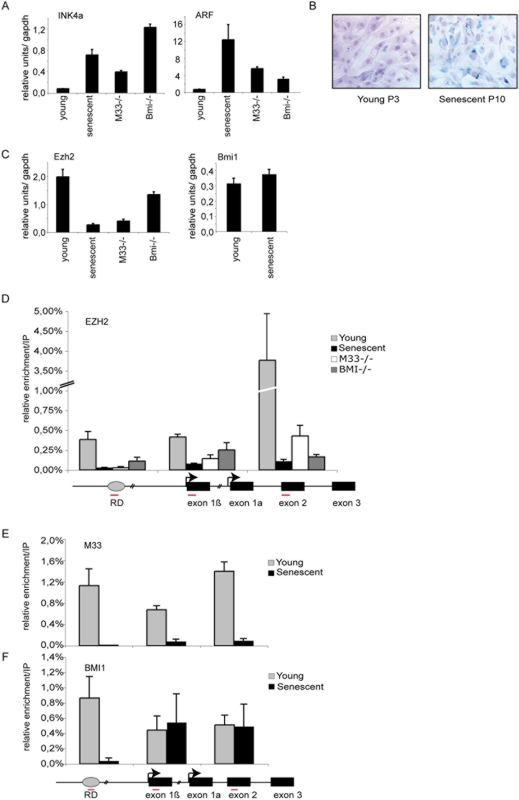
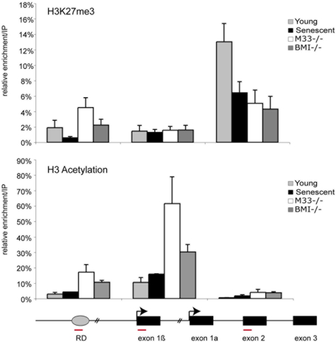
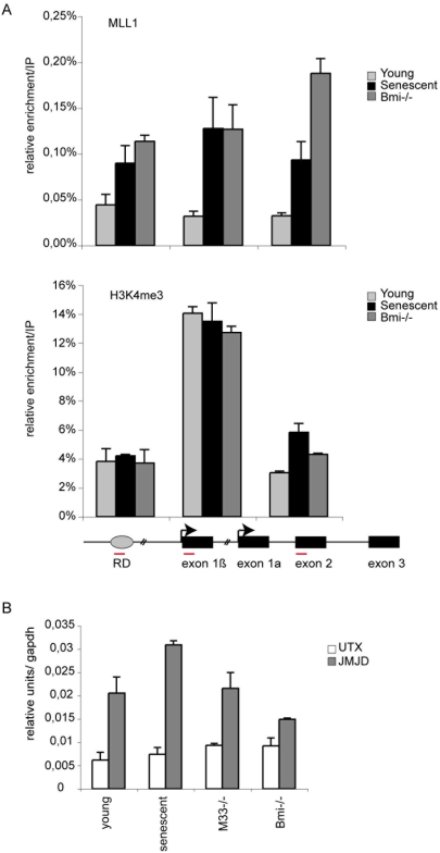
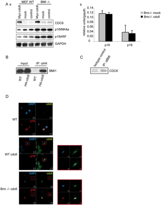
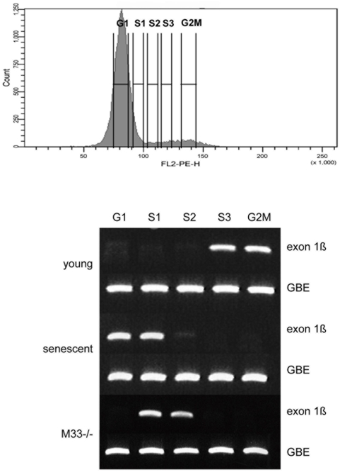
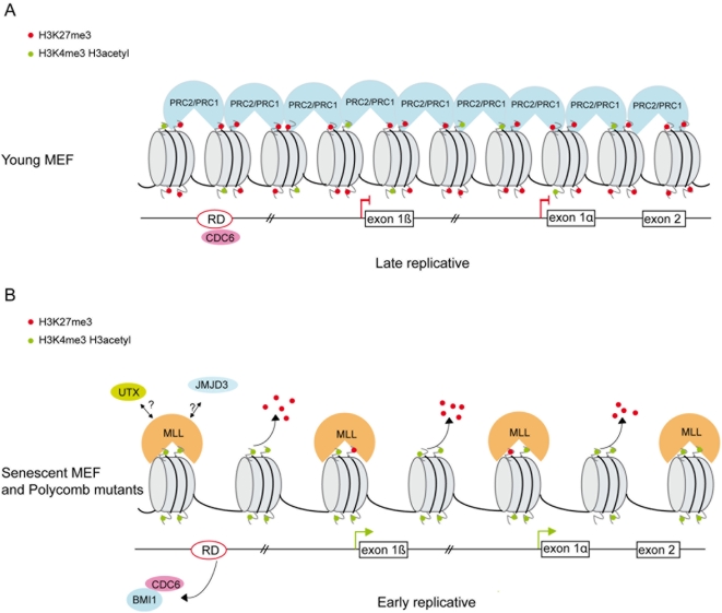
Principal findings: Here we show that in young proliferating embryonic fibroblasts (MEFs) the Polycomb Repressive Complex 2 (PRC2) member EZH2 together with PRC1 members BMI1 and M33 are strongly expressed and localized at the INK4/ARF regulatory domain (RD) identified as a DNA replication origin. When cells enter senescence the binding to RD of both PRC1 and PRC2 complexes is lost leading to a decreased level of histone H3K27 trimethylation (H3K27me3). This loss is accompanied with an increased expression of the histone demethylase Jmjd3 and with the recruitment of the MLL1 protein, and correlates with the expression of the Ink4a/Arf genes. Moreover, we show that the Polycomb protein BMI1 interacts with CDC6, an essential regulator of DNA replication in eukaryotic cells. Finally, we demonstrate that Polycomb proteins and associated epigenetic marks are crucial for the control of the replication timing of the INK4a/ARF locus during senescence.

Conclusions: We identified the replication licencing factor CDC6 as a new partner of the Polycomb group member BMI1. Our results suggest that in young cells Polycomb proteins are recruited to the INK4/ARF locus through CDC6 and the resulting silent locus is replicated during late S-phase. Upon senescence, Jmjd3 is overexpressed and the MLL1 protein is recruited to the locus provoking the dissociation of Polycomb from the INK4/ARF locus, its transcriptional activation and its replication during early S-phase. Together, these results provide a unified model that integrates replication, transcription and epigenetics at the INK4/ARF locus.


Conflict of interest statement

Competing Interests: The authors have declared that no competing interests exist.

Figures

Figure 1
Figure 1. Analysis of Polycomb EZH2, M33 and BMI1 binding at the INK4a/ARF region.
A) qPCR analysis of p16INK4a and p19ARF in young (P3), senescent (P10) and in Bmi1 and M33 mutant MEFs. B) B-galactosidase staining to detect senescent cells at passage 3 (P3) and passage 10 (P10). C) qPCR analysis of the mRNA levels of Polycomb EZH2 and Bmi1 in the indicated cells. D–F) Schematic diagram of the INK4a/ARF locus: amplified regions that were tested in ChIP experiments are indicated by red bars (sequences are given in Table 1). Wild type P3 (young), P10–12 (senescent), M33−/− (P4) and Bmi−/− (P4) MEFs were subjected to ChIP assays using anti EZH2, BMI1 and M33 antibodies. DNA enrichment was calculated as described in Materials and Methods. Bars represent the mean+/−s.d. of quantifications from two to four separate immunoprecipitations analyzed in triplicate.
Figure 2
Figure 2. Loss of EZH2 binding and H3K27me3 methylation at the INK4a/ARF locus during senescence.
P3 proliferating and P10–12 senescent MEFs were subjected to ChIP assays using the indicated antibodies.
Figure 3
Figure 3. Recruitment of MLL1 at the INK4a/ARF locus during senescence.
A) ChIP analysis of the RD element, p19ARF exon 1b and p16/19 shared exon 2 using MLL-c antibody and H3K4me3 antibody. B) qPCR analysis of the mRNA levels of UTX and JMJD3 histone demethylase in the indicated cells.
Figure 4
Figure 4. BMI1 interacts with CDC6 and is required for CDC6 repressing function.
A) Western Blot analysis of Myc-CDC6 Wild type and Bmi1 mutant transduced cells. The antibodies used are indicated. GAPDH antibody is used as a loading control. Ab) quantitative PCR experiment showing p16INK4a and p19ARF expression in Myc-CDC6 transduced Bmi1 mutant cells. B) CDC6 interacts specifically with BMI1: HA immunoprecipitated proteins extracted from HA-CDC6 transfected cells were separated by SDS-PAGE and immunoblotted with a BMI1 antibody. C) anti-Bmi1 immunoprecipitated proteins extracted from wild type thymocytes and immunoblotted with a CDC6 antibody. D) BMI1 is required for INK4a CDC6 mediated repression. Wild type MEFs transfected with Myc-CDC6 were immunostained with a specific antibody against p16INK4a (red) and CDC6 (green) middle panel. Untransfected cells are shown on the upper panel. Bmi1 mutant cells transfected with Myc-CDC6 (green) express high level of p16 (red) (Bottom panel).
Figure 5
Figure 5. INK4a/ARF timing of replication.
PCR based analysis of replication timing of the INK4a/ARF locus (exon 1b). BrdU pulse labeled cells were stained for DNA content with propidium iodide and sorted by flow cytometry into 5 cell cycle fractions (G1, S1, S2, S3 and G2M) according to DNA content. The Gbe D. melanogaster gene provides a control for recovery of BrdU-labeled DNA.
Figure 6
Figure 6. Model for Pc-G and MLL1 proteins in regulation of cellular senescence at the INK4a/ARF locus.
(A) In young proliferating cells, the PRC2 complex is bound at RD and at the INK4a/ARF locus and maintains the levels of H3K27me3. This allows the association of M33 and BMI1-containing PRC1 complex and repression of the INK4a/ARF genes. (B) In senescent or Polycomb mutant cells binding of EZH2 is lost, leading to the disruption of the PRC2 complex, the loss of H3K27me3 and to the recruitment of the MLL1 protein. We propose a model in which Polycomb/MLL1 and JMJD3 epigenetic modifications at the RD element impact the replication timing and the expression of the locus. Moreover, in senescent cells BMI1 binding is specifically lost at the RD element.

Similar articles

Cited by

References

    1. Campisi J. Cancer, aging and cellular senescence. In Vivo. 2000;14:183–188. - PubMed
    1. Dimri GP, Campisi J. Molecular and cell biology of replicative senescence. Cold Spring Harb Symp Quant Biol. 1994;59:67–73. - PubMed
    1. Sherr CJ, DePinho RA. Cellular senescence: mitotic clock or culture shock? Cell. 2000;102:407–410. - PubMed
    1. Core N, Joly F, Boned A, Djabali M. Disruption of E2F signaling suppresses the INK4a-induced proliferative defect in M33-deficient mice. Oncogene. 2004;23:7660–7668. - PubMed
    1. Sasaki M, Ikeda H, Sato Y, Nakanuma Y. Decreased expression of Bmi1 is closely associated with cellular senescence in small bile ducts in primary biliary cirrhosis. Am J Pathol. 2006;169:831–845. - PMC - PubMed

Publication types

MeSH terms