. 2009 Jul;292(7):994-1027.
doi: 10.1002/ar.20916.

Architectonic subdivisions of neocortex in the tree shrew (Tupaia belangeri)

Affiliations

Architectonic subdivisions of neocortex in the tree shrew (Tupaia belangeri)

Peiyan Wong et al. Anat Rec (Hoboken). 2009 Jul.

Abstract

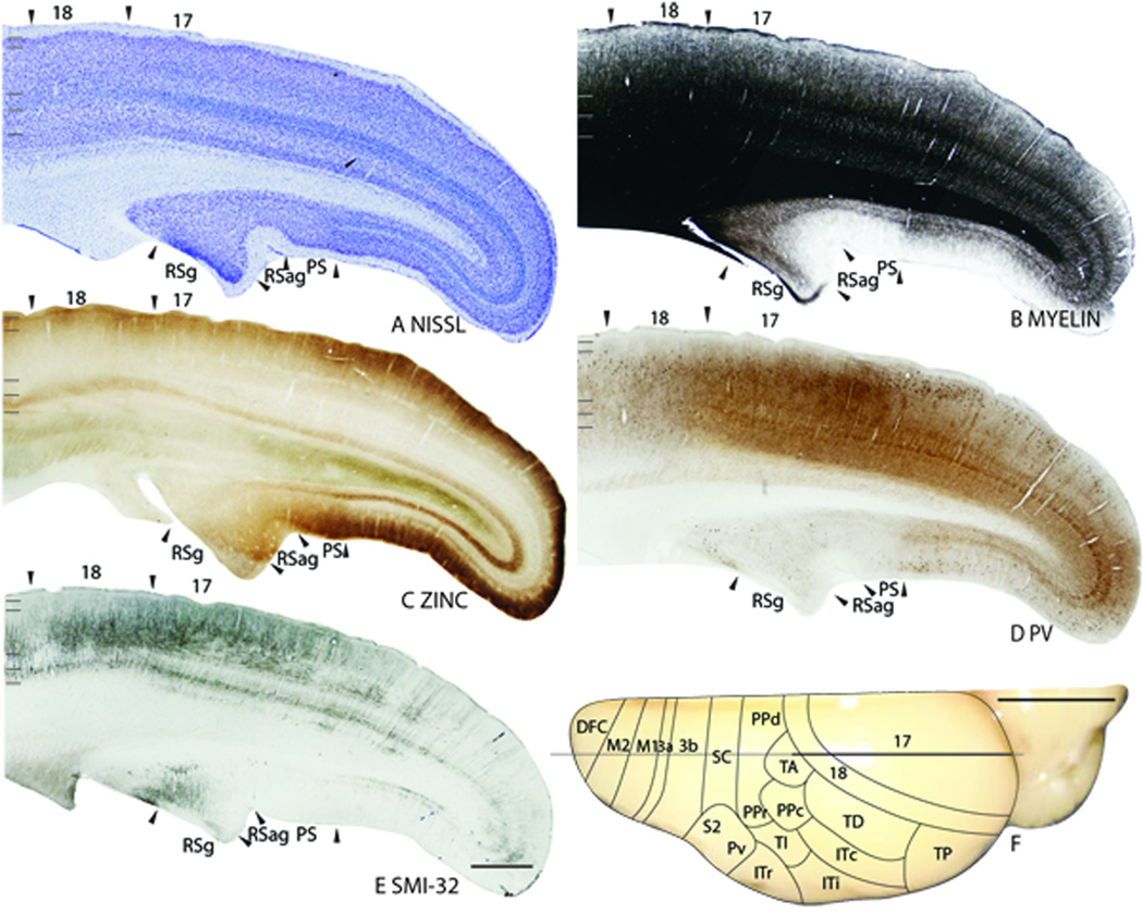
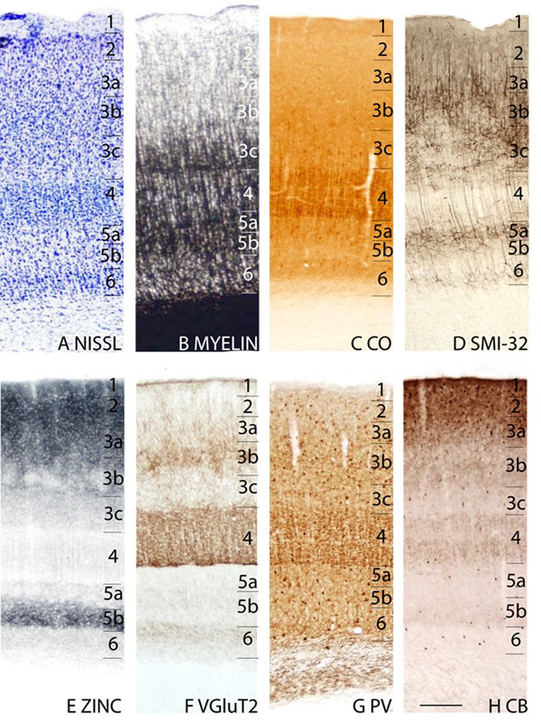
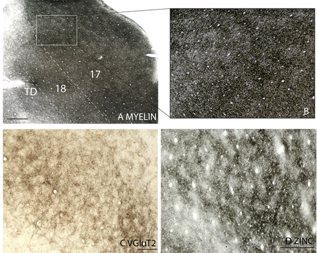
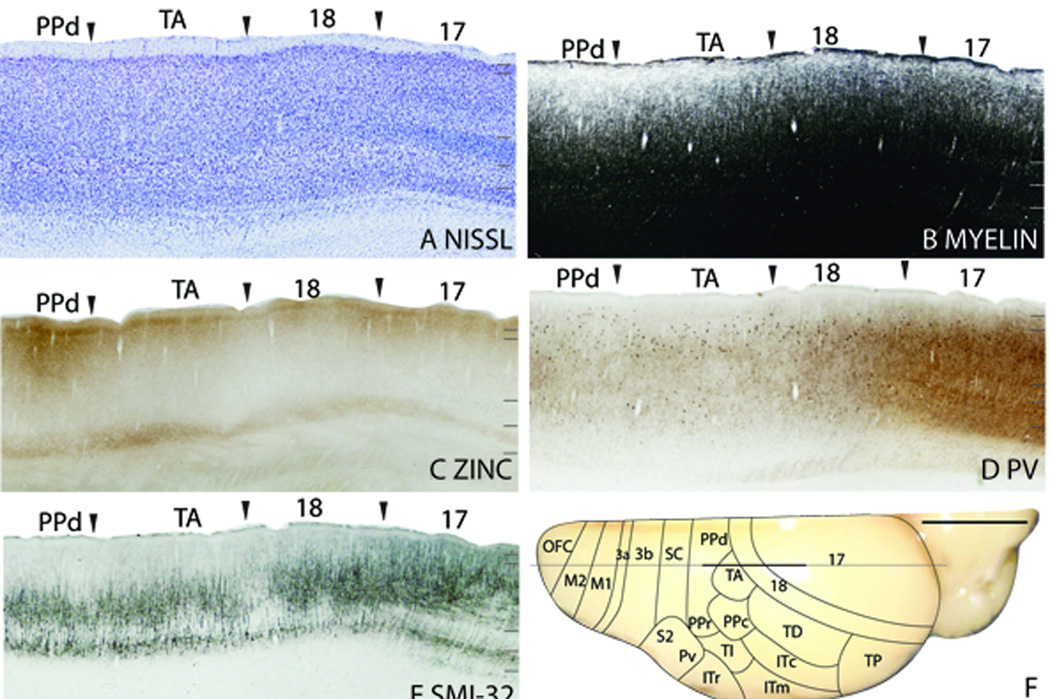
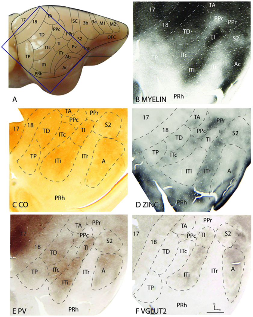
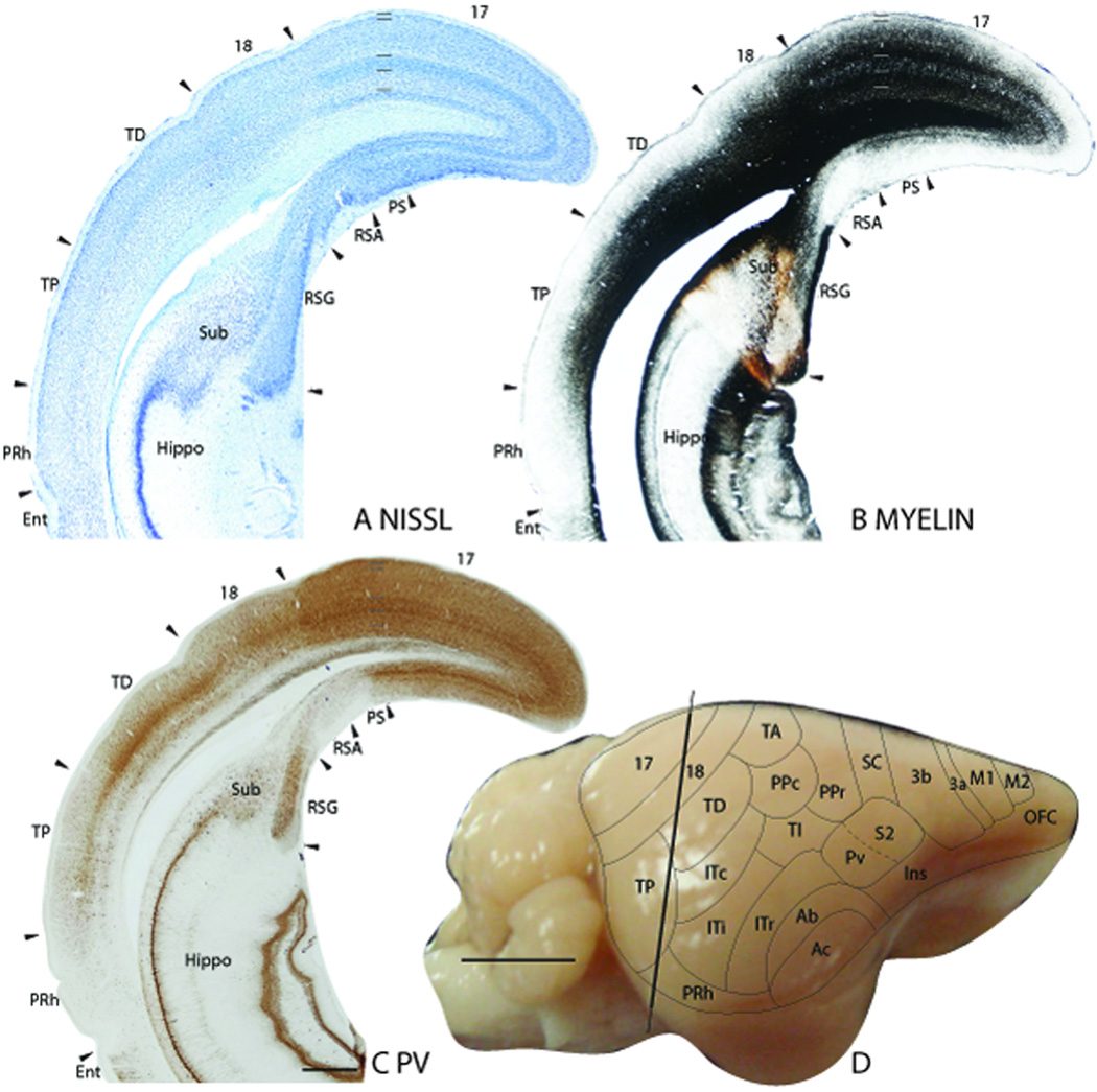
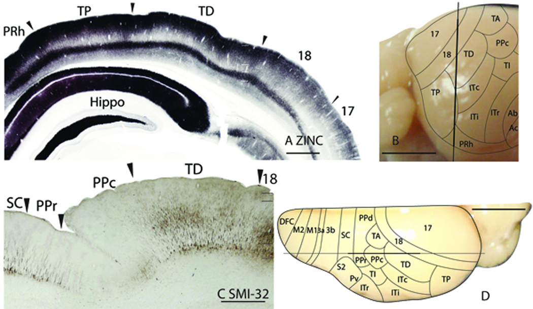
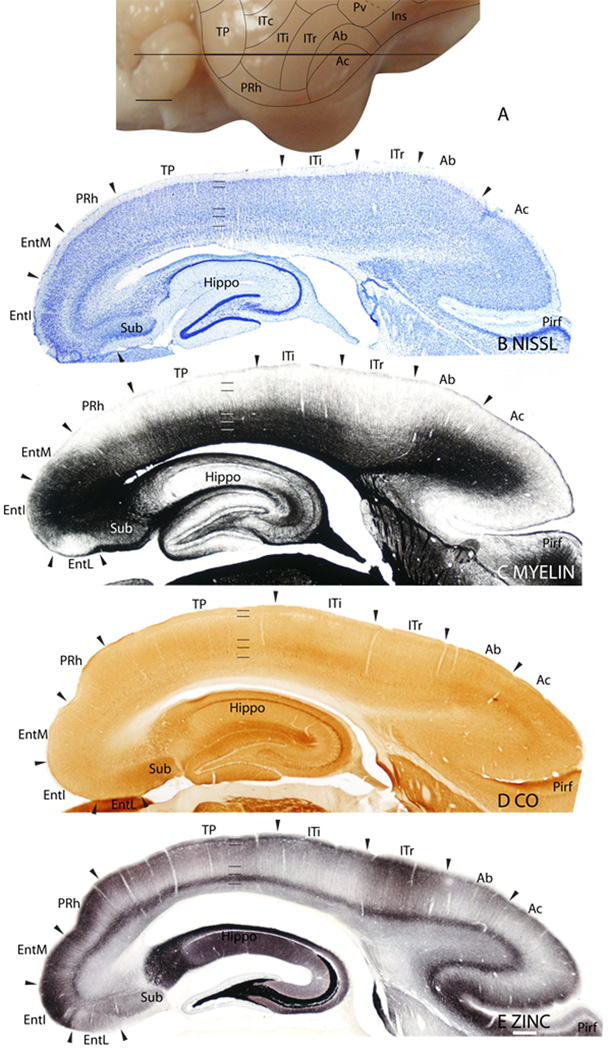
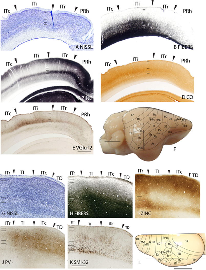
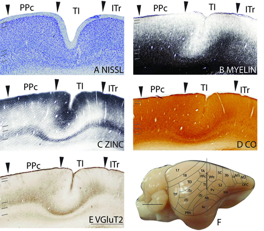
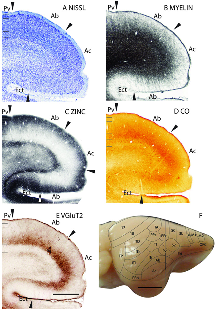
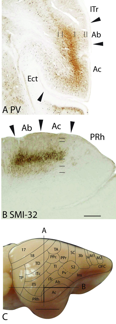
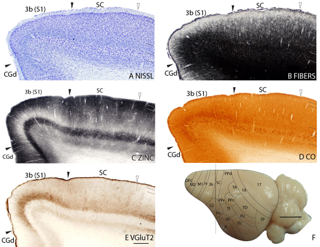
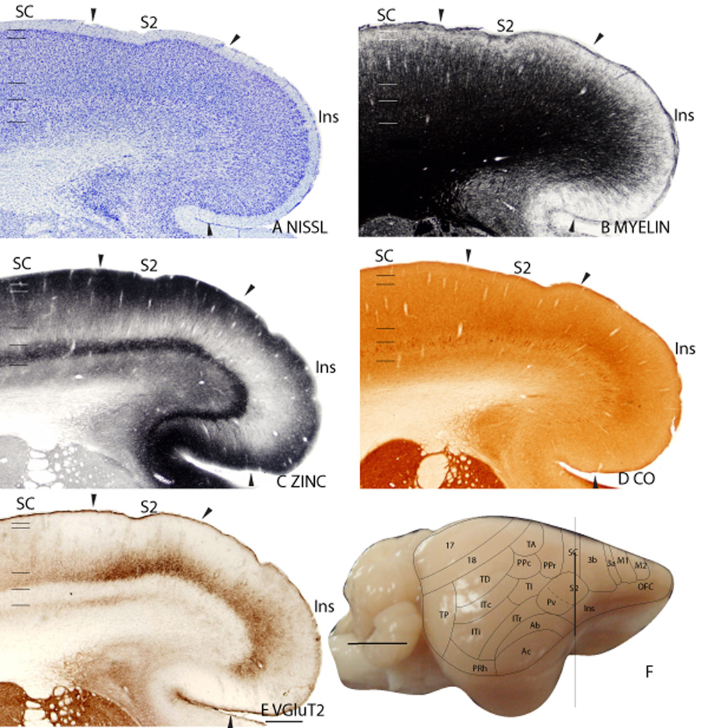
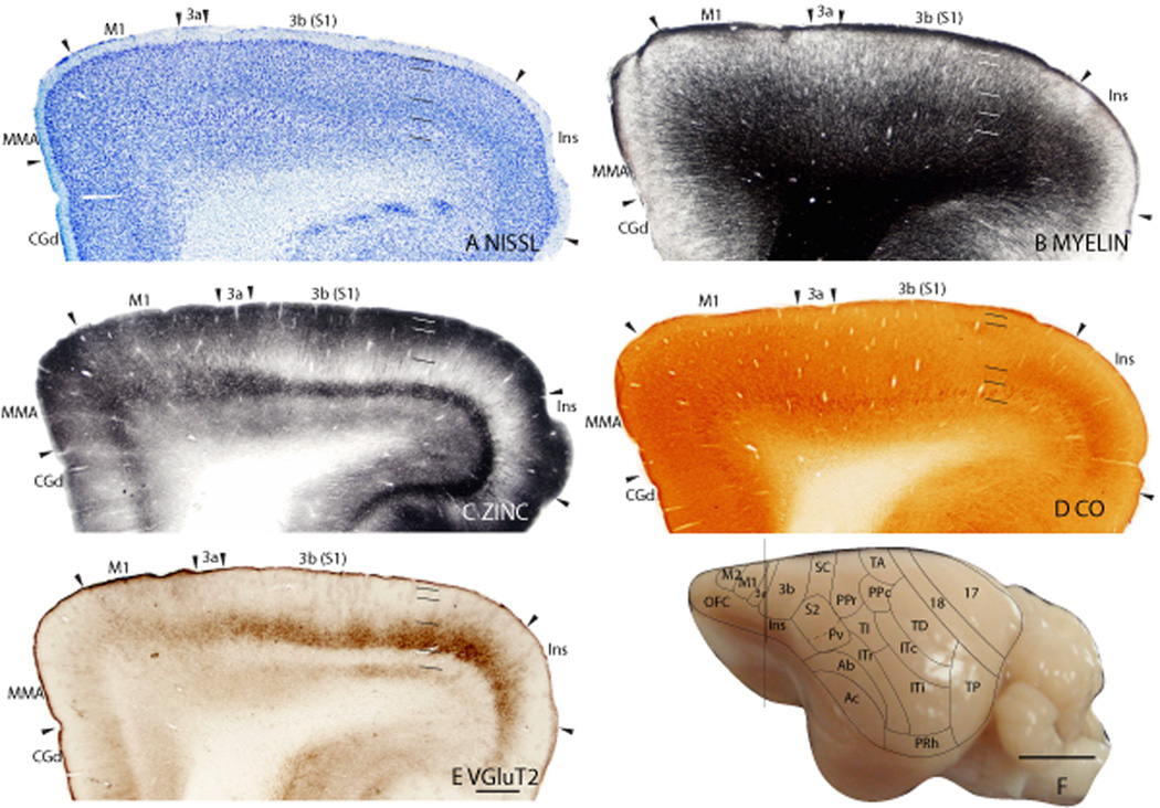
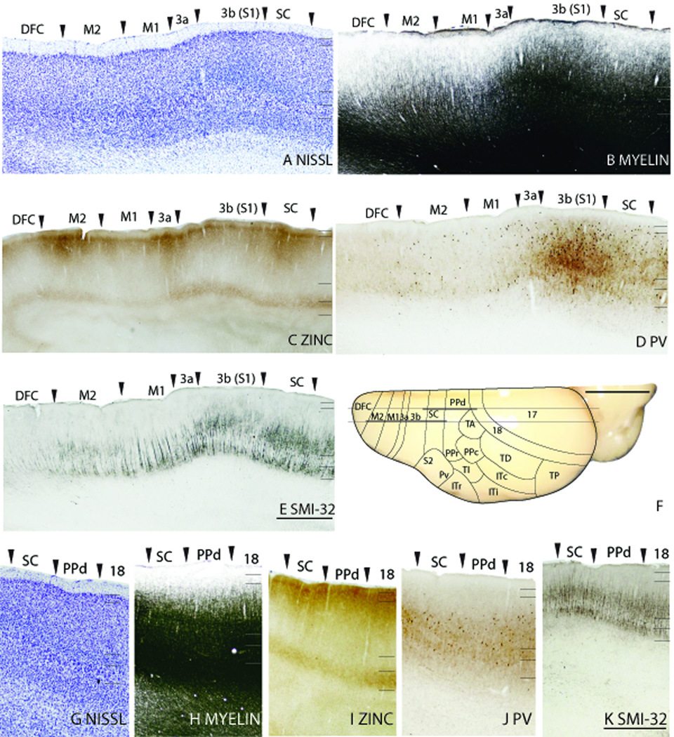
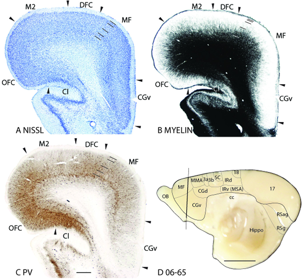
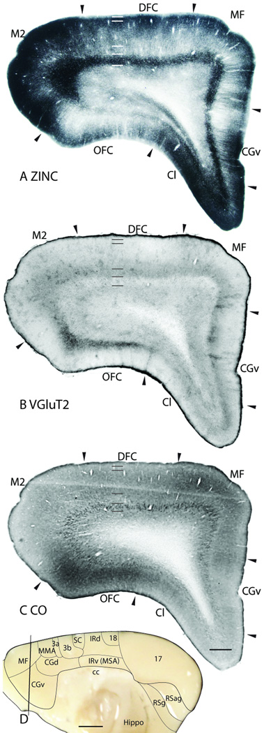
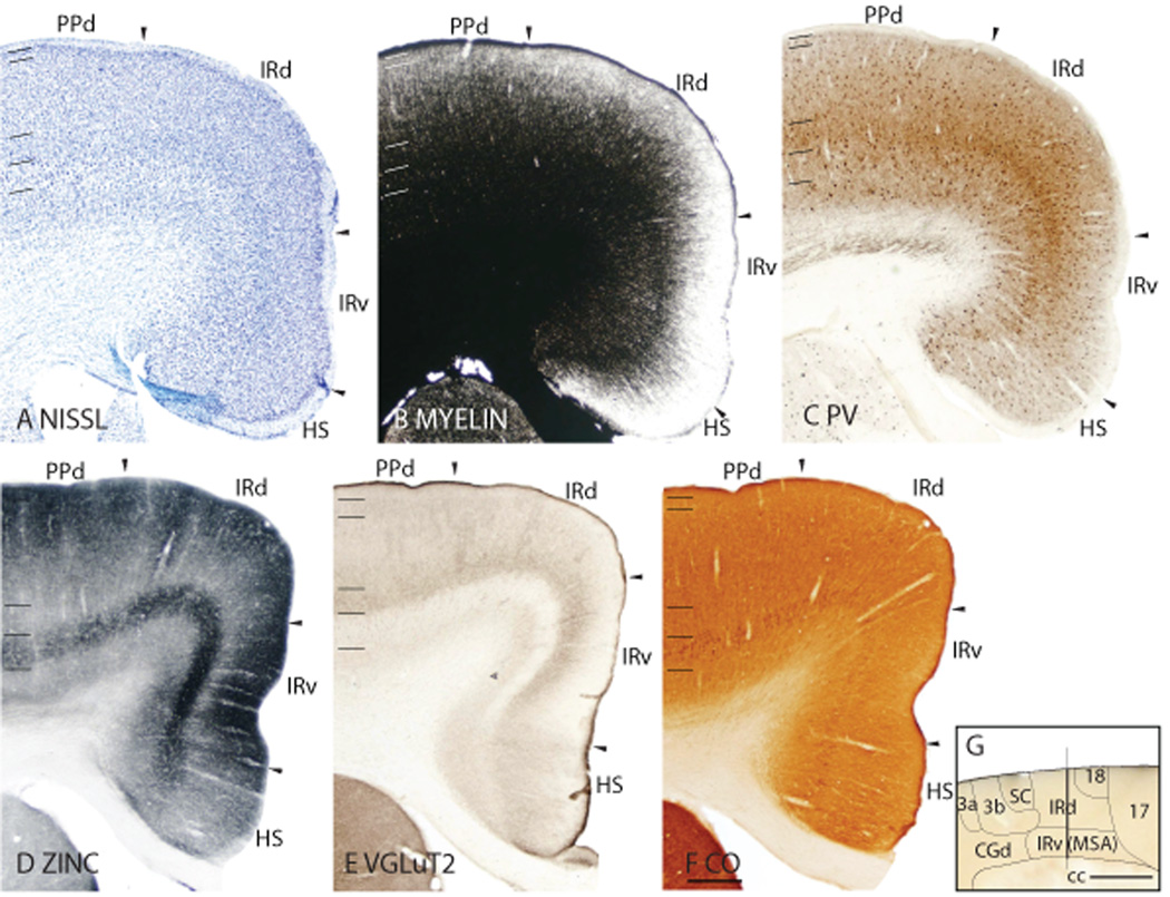
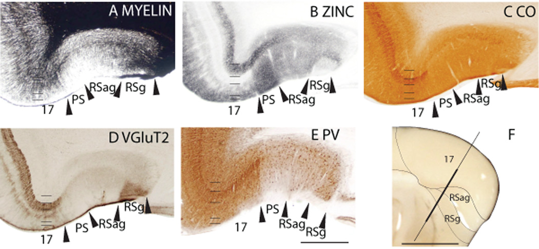
Tree shrews are small mammals that bear some semblance to squirrels, but are actually close relatives of primates. Thus, they have been extensively studied as a model for the early stages of primate evolution. In this study, subdivisions of cortex were reconstructed from brain sections cut in the coronal, sagittal, or horizontal planes, and processed for parvalbumin, SMI-32-immunopositive neurofilament protein epitopes, vesicle glutamate transporter 2 (VGluT2), free ionic zinc, myelin, cytochrome oxidase, and Nissl substance. These different procedures revealed similar boundaries between areas, suggesting the detection of functionally relevant borders and allowed a more precise demarcation of cortical areal boundaries. Primary cortical areas were most clearly revealed by the zinc stain, because of the poor staining of layer 4, as thalamocortical terminations lack free ionic zinc. Area 17 (V1) was especially prominent, as the broad layer 4 was nearly free of zinc stain. However, this feature was less pronounced in primary auditory and somatosensory cortex. In primary sensory areas, thalamocortical terminations in layer 4 densely express VGluT2. Auditory cortex consists of two architectonically distinct subdivisions, a primary core region (Ac), surrounded by a belt region (Ab) that had a slightly less developed koniocellular appearance. Primary motor cortex (M1) was identified by the absence of VGluT2 staining in the poorly developed granular layer 4 and the presence of SMI-32-labeled pyramidal cells in layers 3 and 5. The presence of well-differentiated cortical areas in tree shrews indicates their usefulness in studies of cortical organization and function.


Figures

Fig 1
Fig 1
Architectonic characteristics of visual areas 17, 18 and TD. Coronal sections from occipital cortex were processed for (A) Nissl substance, (B) myelin, (C) synaptic zinc, (D) CO, and (E) vesicle glutamate transporter 2 (VGluT2). The architectonic borders of proposed cortical areas are shown on the dorsal view of the tree shrew brain in panel F. The vertical line on the brain shows the level from which the sections were taken for panels A–E. The thicker portion of the line marks the regions illustrated in panels A–E. Occipital areas 17 and 18 are adopted from Brodmann (1909). TD is the temporal dorsal visual area. Arrowheads on the sections illustrated here and in the following figures mark architectonic boundaries. Short lines on the sections indicate cortical layers 1 to 6. See table 1 for abbreviations for other areas. The scale bar for brain sections (panel E) = 0.5mm. The scale bar on the brain (panel F) = 5mm.
Fig 2
Fig 2
Architectonic characteristics of visual areas and adjoining retrosplenial cortex. Parasagittal sections from occipital cortex were processed for (A) Nissl substance, (B) myelin, (C) synaptic zinc, (D) PV, and (E) neurofilaments with the SMI-32 antibody. The level at which the sagittal sections are taken from is indicated by the horizontal line on the dorsal view of the brain in panel F. The thicker line in panel F marks the regions illustrated in panels A–E. Short lines on the sections indicate the extent of each cortical layers 1 to 6. See table 1 for abbreviations for other areas. The scale bar for brain sections (panel E) = 1mm. The scale bar on the brain (panel F) = 5mm.
Fig 3
Fig 3
The laminar characteristics of area 17 at higher magnification. The sublayers of layer 3 are apparent in the Nissl, synaptic zinc, VGluT2 and CB preparations. Layer 5 has two sublayers, 5a and 5b, that are apparent in Nissl, CO, SMI-32 and zinc preparations. Scale bar = 0.25mm.
Fig 4
Fig 4
Patchy staining pattern of area 17. A. A myelin stained section cut parallel to the surface of an artificially flattened cerebral hemisphere. Dashed lines show the approximate location of the cortical borders. The boxed region in A is shown in B at higher magnification. C and D are from a different case and show the patchy staining pattern of area 17 in VGLuT2 and zinc preparations. Scale bar in panel A = 0.5mm, for panel B and C = 0.5mm, for panel D = 250µm.
Fig 5
Fig 5
Architectonic characteristics of visual and temporal visual areas. The level at which the sagittal sections are taken from is indicated by the horizontal line on the dorsal view of the brain in panel F. The thicker line in panel F marks the regions illustrated in panels A–E. The extent of each cortical layers 1 to 6 is indicated by the short horizontal lines on panels A–E. The scale bar for brain sections (panel E) = 1mm. The scale bar on the brain (panel F) = 5mm.
Fig 6
Fig 6
Architectonic characteristics of temporal and inferior temporal cortex in flattened preparations. A. Architectonic borders of proposed cortical areas are shown on the lateral view of the tree shrew brain. The box indicates the regions that are illustrated in panels B to F. Sections in panels B to F are cut parallel to the surface of an artificially flattened cerebral hemisphere and show the topographic organization of the areas in the temporal and inferior temporal cortex. Scale bar in panel A = 5mm, in panel F = 2mm.
Fig 7
Fig 7
Architectonic characteristics of occipital and temporal visual areas. The location from which the coronal sections are taken from is indicated by the vertical line on the lateral view of the brain in panel D. The thicker portion of the line indicates the region that is illustrated in panels A to C. The scale bar for brain sections (panel C) = 1mm. The scale bar on the brain (panel D) = 5mm.
Fig 8
Fig 8
Architectonic characteristics of the temporal dorsal area. A. A coronal section stained for synaptic zinc. The level at which this section is taken from is indicated by the vertical line on the lateral view of the tree shrew brain. C. A parasagittal section stained for the SMI-32 antibody to reveal neurofilaments. The horizontal line on the dorsal view of the tree shrew brain in D indicates the location from which the section in panel C is taken from. The scale bar for brain sections (panels A and C) = 1mm. The scale bar on the brain (panels B and D) = 5mm.
Fig 9
Fig 9
Architectonic characteristics of the temporal posterior and inferior temporal areas. Cortical areas are shown on a lateral view of the ventral hemisphere in panel A. The thicker portion of the horizontal line across the brain indicates the location of the horizontal brain sections illustrated in panels B to E. The scale bar for brain sections (panel E) = 0.5mm. The scale bar on the brain (panel F) = 5mm.
Fig 10
Fig 10
Architectonic characteristics of the inferior temporal areas. Panels A to E are coronal sections taken from the approximate location indicated by the vertical line on the lateral view of the tree shrew brain in panel F. Panels G to K are parasagittal sections take from the approximate location indicated by the horizontal line on the dorsal view of the tree shrew brain in panel L. The scale bar for brain sections (panels E and K) = 1mm. The scale bar on the brain (panels F and L) = 5mm.
Fig 11
Fig 11
Architectonic characteristics of the termporal intermediate area and adjoining areas. Cortical areas are shown on a lateral view of the right hemisphere in panel F. The thicker part of the vertical line across the brain indicates the location of the coronal brain sections illustrated in panels A to E. The scale bar for brain sections (panel E) = 1mm. The scale bar on the brain (panel F) = 5mm.
Fig 12
Fig 12
Architectonic characteristics of the auditory areas. Cortical areas are shown on a lateral view of the right hemisphere in panel F. The thicker part of the vertical line across the brain indicates the location of the coronal brain sections illustrated in panels A to E. Short horizontal lines on panels A to F indicate the extent of the 6 cortical layers. The scale bar for brain sections (panel E) = 1mm. The scale bar on the brain (panel F) = 5mm.
Fig 13
Fig 13
Architectonic characteristics of the auditory areas in PV and SMI-32 preparations. Cortical areas are shown on a lateral view of the right hemisphere in panel C. The vertical line indicates the location from which the coronal section in panel A is taken, and the horizontal line indicates the location from which the horizontal section in panel B is taken. The scale bar for brain sections (panel B) = 0.5mm. The scale bar on the brain (panel F) = 5mm.
Fig 14
Fig 14
Architectonic characteristics of the primary somatosensory cortex. Cortical areas are shown on a dorsolateral view of the right hemisphere in panel F. The vertical line across the cortex (panel F) indicates the location where the coronal brain sections in panels A to E were obtained. Short horizontal lines on panels A to E indicate the extent of the 6 cortical layers. Solid arrows mark the extent of 3b(S1). Dotted arrows indicate the approximate location of the border between SC and S2. The scale bar for brain sections (panel E) = 0.5mm. The scale bar on the brain (panel F) = 5mm.
Fig 15
Fig 15
Architectonic characteristics of the secondary somatosensory cortex and insular cortex. Cortical areas are shown on a lateral view of the right hemisphere in panel F. The vertical line across the cortex (panel F) indicates the location where the coronal brain sections in panels A to E were obtained. The extent of each cortical layers 1 to 6 is indicated by the short horizontal lines on panels A–E. The scale bar for brain sections (panel E) = 0.5mm. The scale bar on the brain (panel F) = 5mm.
Fig 16
Fig 16
Architectonic characteristics of primary somatosensory and motor areas. Cortical areas are shown on a lateral view of the left hemisphere in panel F. The vertical line across the cortex (panel F) indicates the location where the coronal brain sections in panels A to E were obtained. Short horizontal lines on panels A to E indicate the extent of the 6 cortical layers. The scale bar for brain sections (panel E) = 0.5mm. The scale bar on the brain (panel F) = 5mm.
Fig 17
Fig 17
Architectonic characteristics of somatosensory and motor areas. Cortical areas are shown on a dorsal view of the left hemisphere in panel F. The lateral most horizontal line across the cortex (panel F) indicates the location where the sagittal brain sections in panels A to E were obtained. The horizontal line across the cortex (panel F) that is close to the medial wall indicates the location where the sagittal brain sections in panels G to K were obtained. The scale bar for brain sections (panels E and K) = 1mm. The scale bar on the brain (panel F) = 5mm.
Fig 18
Fig 18
Architectonic characteristics of the medial frontal and cingulate areas. Cortical areas are shown on the medial view of the right hemisphere (panel D). The vertical line across the cortex (panel D) indicates the location where the coronal brain sections in panels A to C were obtained. The extent of each cortical layers 1 to 6 is indicated by the short horizontal lines on panels A to C. The scale bar for brain sections (panel E) = 0.5mm. The scale bar on the brain (panel F) = 5mm.
Fig 19
Fig 19
Architectonic characteristics of the medial frontal and cingulate ventral area. Cortical areas are shown on the medial view of the right hemisphere (panel D). The vertical line across the cortex (panel D) indicates the location where the coronal brain sections in panels A to C were obtained. Short horizontal lines on panels A to E indicate the extent of the 6 cortical layers. The scale bar for brain sections (panel C) = 0.5mm. The scale bar on the brain (panel D) = 2.5mm.
Fig 20
Fig 20
Architectonic characteristics of the infraradiata areas. Cortical areas are shown on the medial view of the right hemisphere (panel G). The vertical line across the cortex (panel G) indicates the location where the coronal brain sections in panels A to F were obtained. The extent of each cortical layers 1 to 6 is indicated by the short horizontal lines on panels A to F. The scale bar for brain sections (panel F) = 0.5mm. The scale bar on the brain (panel G) = 2.5mm.
Fig 21
Fig 21
Architectonic characteristics of the retrosplenial areas. Cortical areas are shown on the medial view of the right caudal hemisphere (panel F). The vertical line across the cortex (panel F) indicates the location where the coronal brain sections in panels A to E were obtained. The scale bar for brain sections (panel E) = 0.5mm. The scale bar on the brain (panel F) = 5mm.

Similar articles

Cited by

References

    1. Abplanalp P. Some subcortical connections of the visual system in tree shrews and squirrels. Brain Behav Evol. 1970;3(1):155–168. - PubMed
    1. Allman JM, Kaas JH. Representation of the visual field in striate and adjoining cortex of the owl monkey (aotus trivirgatus) Brain Res. 1971;35(1):89–106. - PubMed
    1. Augustine JR. The insular lobe in primates including humans. Neurol Res. 1985;7(1):2–10. - PubMed
    1. Barbas H, Pandya DN. Architecture and frontal cortical connections of the premotor cortex (area 6) in the rhesus monkey. J Comp Neurol. 1987;256(2):211–228. - PubMed
    1. Bishop A. Use of the hand in lower primates. In: Buettner-Janusch J, editor. Evolutionary and genetic biology of primates. New York: Academic Press; 1964. pp. 133–225.

Publication types

MeSH terms

LinkOut - more resources