. 2009 Aug;30(8):1167-74.
doi: 10.1002/humu.21017.

Molecular profiling of the "plexinome" in melanoma and pancreatic cancer

Affiliations

Molecular profiling of the "plexinome" in melanoma and pancreatic cancer

Asha Balakrishnan et al. Hum Mutat. 2009 Aug.

Abstract

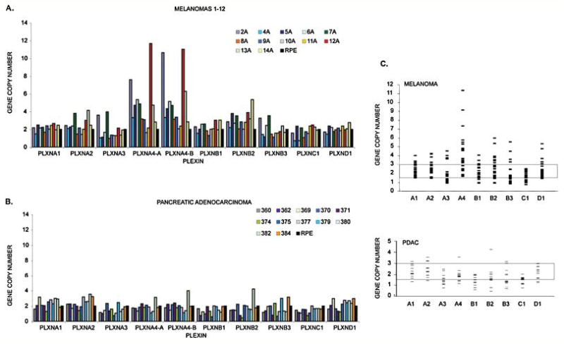
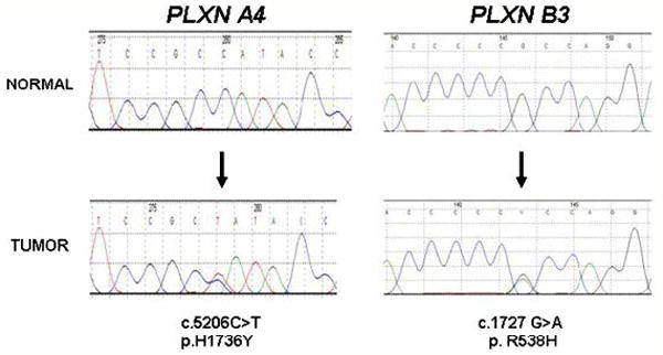
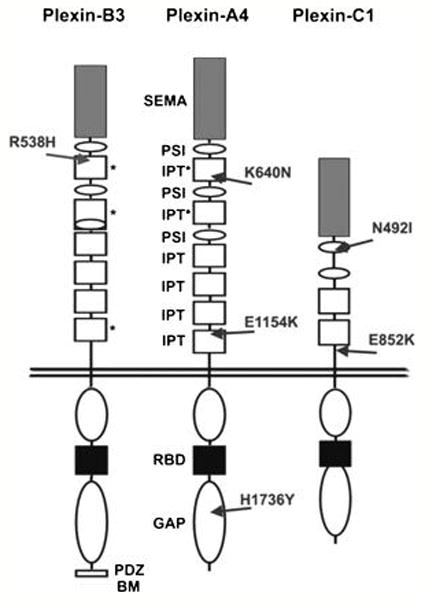
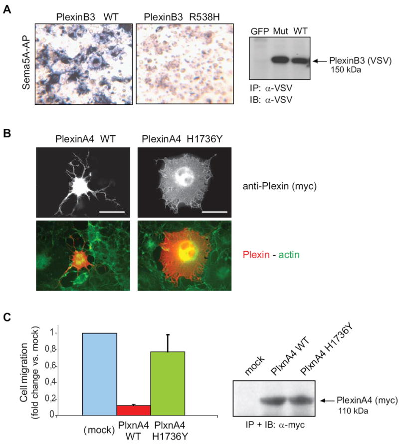
Plexins are transmembrane high-affinity receptors for semaphorins, regulating cell guidance, motility, and invasion. Functional evidences implicate semaphorin signals in cancer progression and metastasis. Yet, it is largely unknown whether plexin genes are genetically altered in human tumors. We performed a comprehensive gene copy analysis and mutational profiling of all nine members of the plexin gene family (plexinome), in melanomas and pancreatic ductal adenocarcinomas (PDACs), which are characterized by high metastatic potential and poor prognosis. Gene copy analysis detected amplification of PLXNA4 in melanomas, whereas copy number losses of multiple plexin genes were seen in PDACs. Somatic mutations were detected in PLXNA4, PLXNB3, and PLXNC1; providing the first evidence that these plexins are mutated in human cancer. Functional assays in cellular models revealed that some of these missense mutations result in loss of plexin function. For instance, c.1613G>A, p.R538H mutation in the extracellular domain of PLXNB3 prevented binding of the ligand Sema5A. Moreover, although PLXNA4 signaling can inhibit tumor cell migration, the mutated c.5206C>T, p.H1736Y allele had lost this activity. Our study is the first systematic analysis of the "plexinome" in human tumors, and indicates that multiple mutated plexins may be involved in cancer progression.


Figures

Figure 1
Figure 1. Plexin gene copy numbers in melanomas and PDACs
We performed Quantitative Real Time PCR with genomic DNA derived from twenty-four melanomas and twelve PDACs, using plexin-specific probes. The figure shows: A, copy number results for the first 12 melanomas (results for the next 12 melanomas are reported in detail in Supp. Figure S1) and B, the twelve PDACs. The nine plexins are indicated on the X-axis (A1 through D1). The Y-axis shows copy numbers after normalization with control DNA (RPE cells). Panel C summarizes the results of copy number analysis of the plexin gene family. Each small bar represents an individual case. Gene copy numbers below 1.5 and above 3 (highlighted range) are normally considered as aberrant.
Figure 2
Figure 2. Somatic mutations of plexin genes in melanoma and PDAC
Chromatograms of two of the seven somatic mutations found in plexin genes in different melanomas and PDAC (other mutations are shown in Supp. Figure S3 and S4). PLXNA4 p.H1736Y mutation was found in melanoma sample 14A, while PLXNB3 p.R538H was identified in PDAC sample 384. In both cases, the lower chromatogram is from the tumor sample, and the upper chromatogram is from the corresponding normal. Arrows indicate the location of missense somatic mutations, and the nucleotide and amino acid alterations are indicated below the traces. Numbers above the sequences are part of the software output.
Figure 3
Figure 3. Schematic representation of plexin domain structure and mutations
The location of novel missense mutations found in human cancer samples are indicated by arrows. In the extracellular portions, grey boxes with “SEMA” indicate semaphorin domains, white ovals with “PSI” indicate Plexin-Semaphorin-Integrin domains (also known as MRS motifs), and white boxes with “IPT” indicate Integrin-Plexin-Transcription-factor domains. Previously unidentified IPT-like domains described in this work are marked by asterisks. In the cytoplasmic portion of the receptors, white ovals indicate the conserved GAP-like regions, the black boxes indicate the Rac-Rnd GTP-ase Binding Domains (RBD), while the C-terminus of PlexinB3 includes a PDZ-domain Binding Motif.
Figure 4
Figure 4. Functional impact of Plexin mutations found in cancers
A, the functional impact of mutation p.R538H in the extracellular domain of PLXNB3 was assessed in COS cells (see Supplementary Methods for details) by challenging the receptor with a soluble form of the specific ligand Sema5A fused to alkaline phosphatase (Sema5A-AP), as previously shown (Artigiani et al., 2004). The comparable expression of the receptor proteins in the cells used for binding assays was confirmed by Western blotting (on the right). The results shown are representative of three independent experiments. The mutated extracellular domain of PLXNB3 has lost the ability to interact with Sema5A. B, the collapsing activity of either wild type or p.H1736Y mutated PLXNA4 was tested in COS cells, by expressing a constitutively active form of the receptor (see Methods), followed by immunofluorescence analysis with anti-Myc-tag antibodies (revealing the plexin) and phalloidin-FITC cell counterstaining (see merged images, at the bottom). Scale bar = 30 μm. The comparable expression of the two proteins was confirmed by Western blotting (on the right). Over 60% of the cells expressing wild-type active PlexinA4 displayed a collapsed phenotype (as shown in representative field), while the mutated plexin had lost this ability (yielding less then 10% collapsed cells). The results shown are representative of three independent experiments. C, wild type and p.H1736 mutated PLXNA4 (as in B) were further expressed in MDA-MB435 human melanoma cells, to investigate the impact of the mutation on the ability of plexin signals to inhibit tumor cell migration (see Methods for experimental details). Spontaneous cell migration was significantly inhibited in cells expressing the wild type activated form of PLXNA4, as expected, whereas cells expressing the mutated receptor were almost unaffected, indicating a functional loss. Protein expression levels were comparable (shown on the right).

Similar articles

Cited by

References

    1. Akbari M, Hansen MD, Halgunset J, Skorpen F, Krokan HE. Low copy number DNA template can render polymerase chain reaction error prone in a sequence-dependent manner. J Mol Diagn. 2005;7:36–39. - PMC - PubMed
    1. Artigiani S, Conrotto P, Fazzari P, Gilestro GF, Barberis D, Giordano S, Comoglio PM, Tamagnone L. Plexin-B3 is a functional receptor for semaphorin 5A. EMBO Rep. 2004;5:710–714. - PMC - PubMed
    1. Balakrishnan A, Bleeker FE, Lamba S, Rodolfo M, Daniotti M, Scarpa A, van Tilborg AA, Leenstra S, Zanon C, Bardelli A. Novel somatic and germline mutations in cancer candidate genes in glioblastoma, melanoma, and pancreatic carcinoma. Cancer Res. 2007;67:3545–3550. - PubMed
    1. Balch CM, Soong SJ, Gershenwald JE, Thompson JF, Reintgen DS, Cascinelli N, Urist M, McMasters KM, Ross MI, Kirkwood JM, Atkins MB, Thompson JA, Coit DG, Byrd D, Desmond R, Zhang Y, Liu PY, Lyman GH, Morabito A. Prognostic factors analysis of 17,600 melanoma patients: validation of the American Joint Committee on Cancer melanoma staging system. J Clin Oncol. 2001;19:3622–3634. - PubMed
    1. Barberis D, Artigiani S, Casazza A, Corso S, Giordano S, Love CA, Jones EY, Comoglio PM, Tamagnone L. Plexin signaling hampers integrin-based adhesion, leading to Rho-kinase independent cell rounding, and inhibiting lamellipodia extension and cell motility. Faseb J. 2004;18:592–594. - PubMed

Publication types

Substances