Review
. 2009 May;136(6):1965-78.
doi: 10.1053/j.gastro.2008.12.071.

The gastrointestinal tract and AIDS pathogenesis

Affiliations
Review

The gastrointestinal tract and AIDS pathogenesis

Andrew A Lackner et al. Gastroenterology. 2009 May.

Abstract

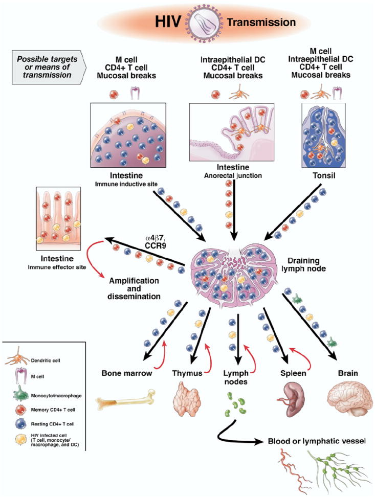
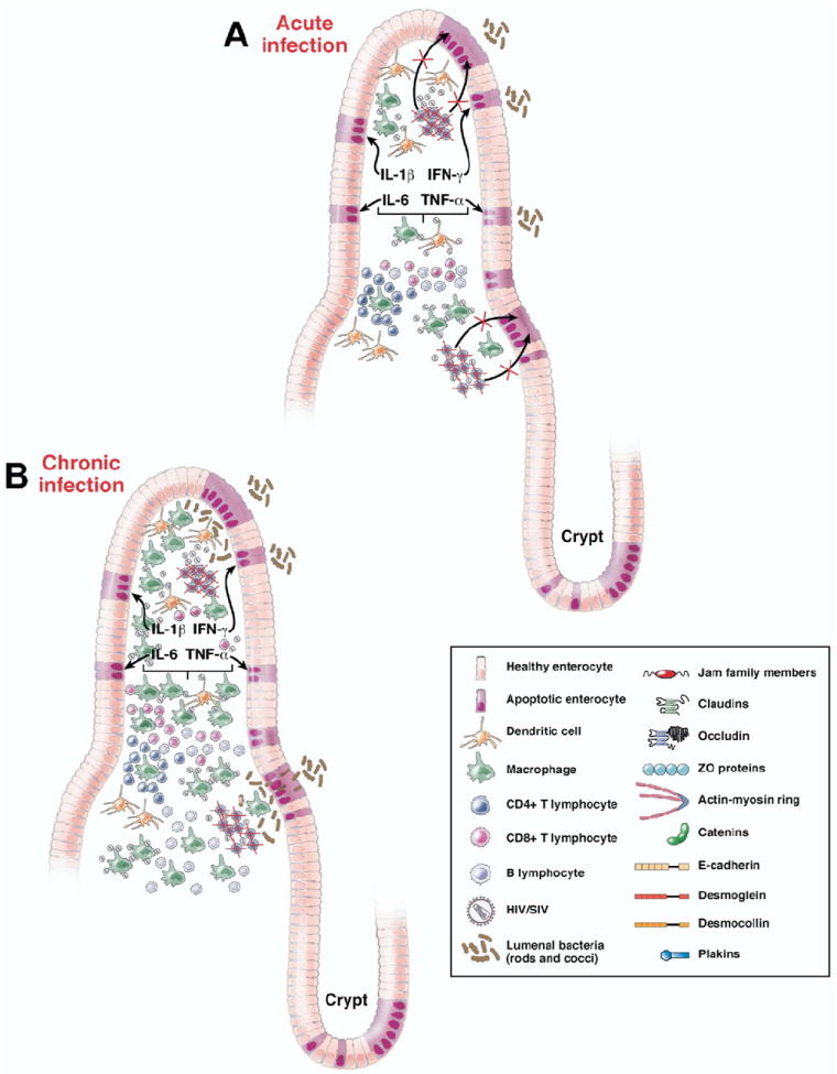
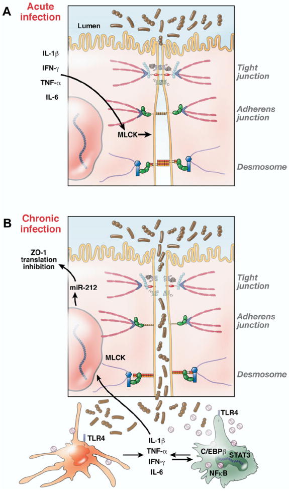
Gastrointestinal disease has been recognized as a major manifestation of human immunodeficiency virus infection since the earliest recognition of acquired immunodeficiency syndrome (AIDS). Originally, these disease manifestations were considered to be sequelae of the immune destruction that characterizes AIDS rather than being central to the pathogenesis of AIDS. Over time, it has become clear that the mucosal immune system in general and the intestinal immune system in particular are central to the pathogenesis of AIDS, with most of the critical events (eg, transmission, viral amplification, CD4+ T-cell destruction) occurring in the gastrointestinal tract. Compared with peripheral blood, these tissues are not easily accessible for analysis and have only begun to be examined in detail recently. In addition, although the resulting disease can progress over years, many critical events happen within the first few weeks of infection, when most patients are unaware that they are infected. Moreover, breakdown of the mucosal barrier and resulting microbial translocation are believed to be major drivers of AIDS progression. In this review, we focus on the interaction between primate lentiviruses and the gastrointestinal tract and discuss how this interaction promotes the pathogenesis of AIDS and drives immune dysfunction and progression to AIDS. This article draws extensively on work done in the nonhuman primate model of AIDS to fill gaps in our understanding of AIDS in humans.


Conflict of interest statement

Conflicts of interest

The authors disclose no conflicts.

Figures

Figure 1
Figure 1
Most infections with HIV occur via mucosal surfaces. In the gastrointestinal tract, this involves the anorectal mucosa and possibly tonsil. In addition to mucosal breaks that facilitate transmission, there are several other cellular targets for mucosal transmission. This includes M cells in intestine and tonsil associated with immune inductive sites and intraepithelial DCs, particularly in the anorectal mucosa. Once the virus crosses the mucosa, it can encounter numerous CD4+ cells, but most T cells in immune inductive sites are resting and are not likely to support infection. In contrast, a high percentage of memory CD4+ T cells that express CCR5 are present in mucosal immune effector sites, which serve as a major site for amplification of the initial infection. Infected cells and virus reach these sites by taking advantage of normal trafficking patterns facilitated by molecules such as α4β7 and CCR9. Regional draining lymph nodes serve as a nexus where infected mucosal homing lymphocytes encounter systemic lymphocytes and disseminate the infection throughout the body.
Figure 2
Figure 2
Schematic representation of pathological changes underlying intestinal epithelial barrier disruption during HIV/SIV infection. (A) Acute HIV/SIV infection depletes the mucosa of CD4+CCR5 effector memory T cells and Th17 cells, leading to disruption of the bidirectional communication that links the intestinal epithelium with the mucosal immune system necessary for the growth, maintenance, and renewal of epithelial cells and leading to increased epithelial cell apoptosis. (B) During chronic HIV/SIV infection, in addition to the continual destruction of CD4+CCR5 effector memory T cells and Th17 cells there is increased infiltration of inflammatory cells into the lamina propria leading to more proinflammatory cytokine production and widespread distribution of the intestinal epithelial barrier facilitating translocation of lumenal bacteria.
Figure 3
Figure 3
Schematic representation of possible molecular mechanisms underlying intestinal epithelial barrier disruption during HIV/SIV infection. (A) Normal intestinal mucosal barrier integrity is maintained by junctional complexes between intestinal epithelial cells (tight junctions, desmosomes, etc). Proinflammatory cytokines (eg, tumor necrosis factor α, IL-1β, interferon gamma) produced by DCs and macrophages in response to local viral replication can also exacerbate epithelial injury by activating factors such as MLCK, which can alter the structure and integrity of tight junctions. (B) As infection progresses, loss of epithelial integrity leads to increased translocation of luminal bacteria and bacterial products that in turn locally activate innate immune cells (eg, DCs and macrophages) to produce more proinflammatory cytokines, thus creating a localized inflammatory environment. Proinflammatory cytokines also activate a variety of immune cells, including CD4+ T cells, which increase the ability of these cells to support viral replication. The underlying molecular mechanism of macrophage activation is partly evident from the constitutive expression of STAT3 and C/EBPβ, proinflammatory transcription factors activated by IL-6 and interferon gamma. Whereas STAT3 is antiapoptotic, C/EBPβ can transactivate the HIV/SIV LTR, resulting in persistent inflammation, immune activation, and increased virus production. Select micro-RNAs such as miR-212, which is up-regulated in the intestine during inflammation, can also contribute to epithelial barrier breakdown by down-regulating the expression of the tight junction protein ZO-1.

References

    1. Alkhatib G, Combadiere C, Broder CC, et al. CC CKR5: A RANTES, MIP-1α, MIP-1β receptor as a fusion cofactor for macrophage-tropic HIV-1. Science. 1996;272:1955–1958. - PubMed
    1. Moore JP, Kitchen SG, Pugach P, et al. The CCR5 and CXCR4 coreceptors—central to understanding the transmission and pathogenesis of human immunodeficiency virus type 1 infection. AIDS Res Hum Retroviruses. 2004;20:111–126. - PubMed
    1. Koff WC, Johnson PR, Watkins DI, et al. HIV vaccine design: insights from live attenuated SIV vaccines. Nat Immunol. 2006;7:19–23. - PubMed
    1. Picker LJ, Watkins DI. HIV pathogenesis: the first cut is the deepest. Nat Immunol. 2005;6:430–432. - PubMed
    1. Veazey RS, DeMaria M, Chalifoux LV, et al. The gastrointestinal tract as a major site of CD4+ T cell depletion and viral replication in SIV infection. Science. 1998;280:427–431. - PubMed

Publication types

MeSH terms

Substances