. 2009 May 24:10:242.
doi: 10.1186/1471-2164-10-242.

Gene expression during Drosophila melanogaster egg development before and after reproductive diapause

Affiliations

Gene expression during Drosophila melanogaster egg development before and after reproductive diapause

Dean A Baker et al. BMC Genomics. .

Abstract

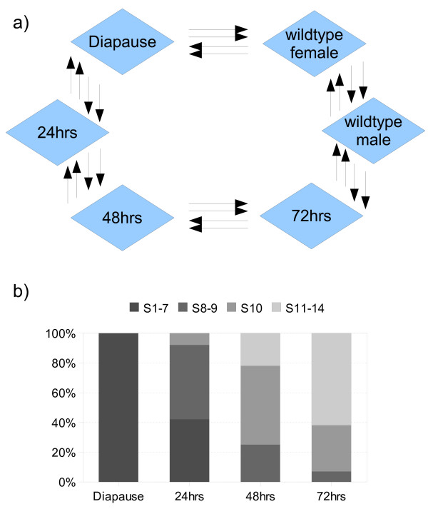
Background: Despite the importance of egg development to the female life cycle in Drosophila, global patterns of gene expression have not been examined in detail, primarily due to the difficulty in isolating synchronised developmental stages in sufficient quantities for gene expression profiling. Entry into vitellogenesis is a key stage of oogenesis and by forcing females into reproductive diapause we are able to arrest oogenesis at the pre-vitellogenic stages. Releasing females from diapause allows collection of relatively synchronous developing egg populations and an investigation of some of the transcriptional dynamics apparent before and after reproductive diapause.

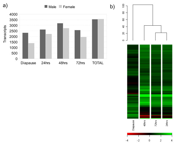
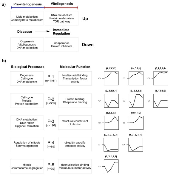
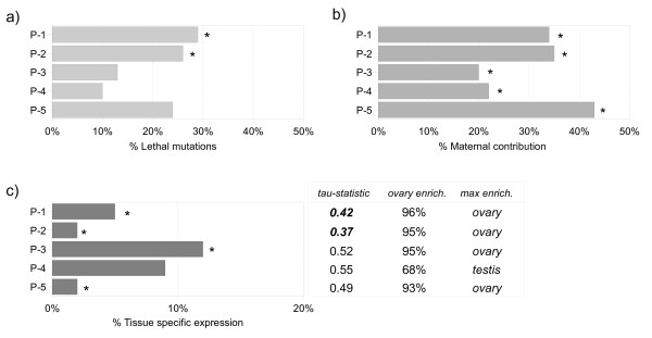
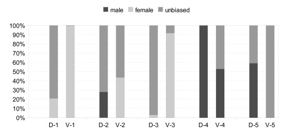
Results: Focusing on gender-biased transcription, we identified mechanisms of egg development suppressed during reproductive dormancy as well as other molecular changes unique to the diapausing female. A microarray based analysis generated a set of 3565 transcripts with at least 2-fold greater expression in females as compared to control males, 1392 such changes were biased during reproductive dormancy. In addition, we also detect 1922 up-regulated transcriptional changes after entry into vitellogenesis, which were classified into discrete blocks of co-expression. We discuss some of the regulatory aspects apparent after re-initiation of egg development, exploring the underlying functions, maternal contribution and evolutionary conservation of co-expression patterns involved in egg production.

Conclusion: Although much of the work we present is descriptive, fundamental aspects of egg development and gender-biased transcription can be derived from our time-series experiment. We believe that our dataset will facilitate further exploration of the developmental and evolutionary characteristics of oogenesis as well as the nature of reproductive arrest in Drosophila.


Figures

Figure 1
Figure 1
Experimental design & ovary dissection. a) Experimental design. Each arrow represents a separate biological replicate and indicates the Cy3 to Cy5 direction of dye swaps performed. b) Ovary dissection. Ovary dissection through the oogenic cycle and the relative proportion of ovarioles with maximum development in Stage 1–14 (n = 15).
Figure 2
Figure 2
Global patterns of gender-biased expression. a) The ratio of gender-biased expression through the oogenic cycle. The Y-axis plots the number of transcripts biased in each sex according to each time point we examined. b) Hierarchical clustering of gender-biased similarity. Genes and samples were clustered across the female-biased dataset with centred gene expression data using Euclidean Distance to calculate similarity.
Figure 3
Figure 3
Gene ontology summary and co-expression profiles. a) Gene ontology summary of diapause and early vitellogenic regulation, featuring the highest over-represented Biological Processes annotations within each group. b) Gene ontology summary and co-expression clusters of oogenic biased expression. Boxes feature the highest over-represented Biological Processes and Molecular Function annotations within each expression profile (P1-P5). The total number of over-represented functional annotations for each cluster are provided [see Additional file 16, 17, 18, 19, 20]. Clusters were generated with Short Time-series Expression Miner (STEM, [56,57]). At each time-step genes were considered up or down regulated when compared to male controls. Expression profiles with a correlation of at least 0.8 were clustered together, generating five groups [see Additional file 21, 23]. Each cluster shown is over-represented at a P-value < 0.05 in permutation tests. Numbers in brackets represent the relative gene expression changes to which profiles were clustered at each point in the time series.
Figure 4
Figure 4
Database summary of the primary oogenesis regulated profiles. a) Lethal mutations. Proportion of genes annotated as 'Phenotypic class: Lethal'. b) Maternal contributions. Proportion of genes with maternal expression according to Hooper et al., 2007 [3]. c) Tissue-Specific. The number of genes with expression in a single tissue according to the FlyAtlas database. tau-statistic: 1= Specific; 0 = Ubiquitous (See Methods for further detail). Ovary Enrichment: The percentage of genes with higher ovary expression compared to whole-organism expression. Most Enriched Tissue: Tissue with the highest number of enriched expression versus whole-organism estimates. Expected values were determined from genome-wide estimates. Asterisks or numbers highlighted in bold were significant at the P < 0.05 level.
Figure 5
Figure 5
Gender-biased expression ratios within co-expression clusters. Expression profiles (1–5) before (D) and after (V) entry into vitellogenesis are shown. Gender-biased expression was calculated as genes with a 2-fold greater level of expression in either sex (q < 0.05), while unbiased expression was calculated as the remainder.

References

    1. Arbeitman MN, Furlong EE, Imam F, Johnson E, Null BH, Baker BS, Krasnow MA, Scott MP, Davis RW, White KP. Gene expression during the life cycle of Drosophila melanogaster. Science. 2002;297:2270–2275. - PubMed
    1. Stolc V, Gauhar Z, Mason C, Halasz G, van Batenburg MF, Rifkin SA, Hua S, Herreman T, Tongprasit W, Barbano PE, Bussemaker HJ, White KP. A gene expression map for the euchromatic genome of Drosophila melanogaster. Science. 2004;306:655–660. - PubMed
    1. Hooper SD, Boué S, Krause R, Jensen LJ, Mason CE, Ghanim M, White KP, Furlong EE, Bork P. Identification of tightly regulated groups of genes during Drosophila melanogaster embryogenesis. Mol Syst Biol. 2007;3:72. - PMC - PubMed
    1. Spradling AC. Developmental Genetics of Oogenesis. In: Bate M, Arias AM, editor. The Development of Drosophila melanogaster. Cold Spring Harbor Laboratory Press; 1993. pp. 1–70.
    1. Mahowald AP, Kambysellis MP. Oogenesis. In: Ashburner M, Wright TRW, editor. The Genetics and Biology of Drosophila. 2. Academic; 1980. pp. 141–224.

Publication types

LinkOut - more resources