

Antegrade percutaneous closure of membranous ventricular septal defect using X-ray fused with magnetic resonance imaging
- PMID: 19463430
- PMCID: PMC2698297
- DOI: 10.1016/j.jcin.2008.09.014
Antegrade percutaneous closure of membranous ventricular septal defect using X-ray fused with magnetic resonance imaging
Abstract
Objectives: We hypothesized that X-ray fused with magnetic resonance imaging (XFM) roadmaps might permit direct antegrade crossing and delivery of a ventricular septal defect (VSD) closure device and thereby reduce procedure time and radiation exposure.
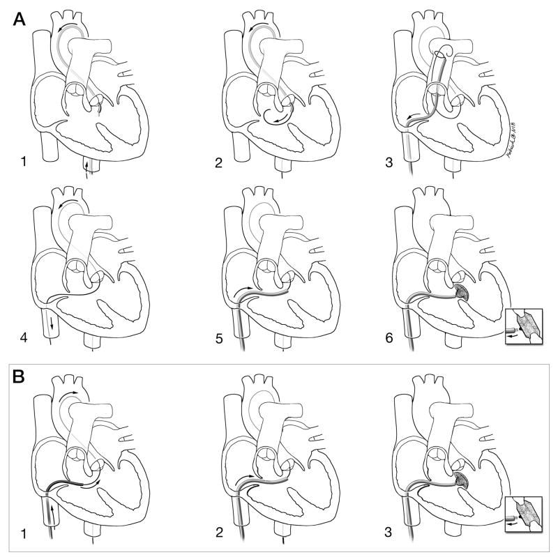
Background: Percutaneous device closure of membranous VSD is cumbersome and time-consuming. The procedure requires crossing the defect retrograde, snaring and exteriorizing a guidewire to form an arteriovenous loop, then delivering antegrade a sheath and closure device.
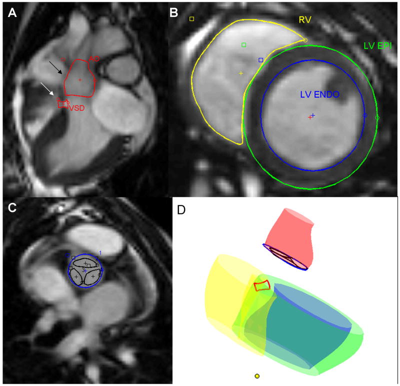
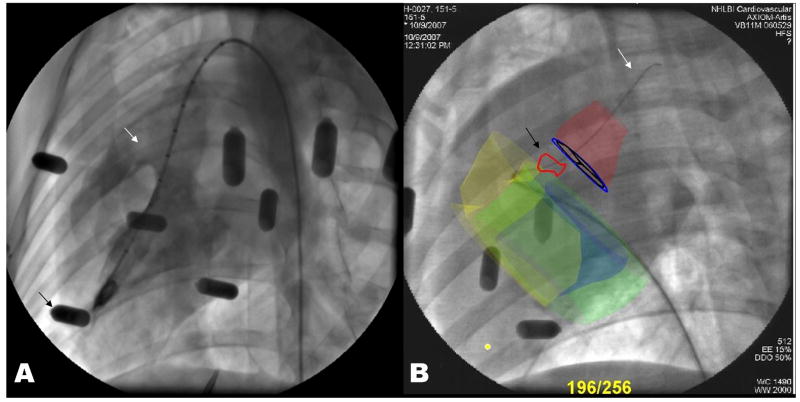
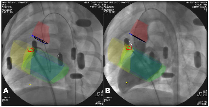
Methods: Magnetic resonance imaging roadmaps of cardiac structures were obtained from miniature swine with spontaneous VSD and registered with live X-ray using external fiducial markers. We compared antegrade XFM-guided VSD crossing with conventional retrograde X-ray-guided crossing for repair.
Results: Antegrade XFM crossing was successful in all animals. Compared with retrograde X-ray, antegrade XFM was associated with shorter time to crossing (167 +/- 103 s vs. 284 +/- 61 s; p = 0.025), shorter time to sheath delivery (71 +/- 32 s vs. 366 +/- 145 s; p = 0.001), shorter fluoroscopy time (158 +/- 95 s vs. 390 +/- 137 s; p = 0.003), and reduced radiation dose-area product (2,394 +/- 1,522 mG.m(2) vs. 4,865 +/- 1,759 mG.m(2); p = 0.016).
Conclusions: XFM facilitates antegrade access to membranous VSD from the right ventricle in swine. The simplified procedure is faster and reduces radiation exposure compared with the conventional retrograde approach.
Figures
Similar articles
-
Directly ventricular septal defect closure without using arteriovenous wire loop: Our adult case series using transarterial retrograde approach.Anatol J Cardiol. 2017 Jun;17(6):461-468. doi: 10.14744/AnatolJCardiol.2017.7507. Epub 2017 Feb 21. Anatol J Cardiol. 2017. PMID: 28315566 Free PMC article.
-
Retrograde closure of perimembranous ventricular septal defect using muscular ventricular septal occluder: a single-center experience of a novel technique.Pediatr Cardiol. 2015 Jan;36(1):106-10. doi: 10.1007/s00246-014-0971-x. Epub 2014 Aug 20. Pediatr Cardiol. 2015. PMID: 25139246
-
Closed-chest transthoracic magnetic resonance imaging-guided ventricular septal defect closure in swine.JACC Cardiovasc Interv. 2011 Dec;4(12):1326-34. doi: 10.1016/j.jcin.2011.09.012. JACC Cardiovasc Interv. 2011. PMID: 22192373 Free PMC article.
-
Retrograde approach for device closure of muscular ventricular septal defects in children and adolescents, using the Amplatzer muscular ventricular septal defect occluder.Pediatr Cardiol. 2006 Nov-Dec;27(6):720-8. doi: 10.1007/s00246-006-1365-5. Epub 2006 Nov 7. Pediatr Cardiol. 2006. PMID: 17091325
-
Percutaneous closure of ventricular septal defects. State of the art.J Cardiovasc Med (Hagerstown). 2007 Jan;8(1):39-45. doi: 10.2459/01.JCM.0000247434.59451.d7. J Cardiovasc Med (Hagerstown). 2007. PMID: 17255815 Review.
Cited by
-
The Future of Paediatric Heart Interventions: Where Will We Be in 2030?Curr Cardiol Rep. 2020 Oct 9;22(12):158. doi: 10.1007/s11886-020-01404-z. Curr Cardiol Rep. 2020. PMID: 33037461 Free PMC article. Review.
-
Roadmaps show the way: coregistration to enhance structural heart interventions.Catheter Cardiovasc Interv. 2013 Sep 1;82(3):443-4. doi: 10.1002/ccd.25115. Catheter Cardiovasc Interv. 2013. PMID: 24038978 Free PMC article. No abstract available.
-
MRI Catheterization: Ready for Broad Adoption.Pediatr Cardiol. 2020 Mar;41(3):503-513. doi: 10.1007/s00246-020-02301-6. Epub 2020 Mar 20. Pediatr Cardiol. 2020. PMID: 32198594 Free PMC article. Review.
-
Interventional cardiovascular magnetic resonance: still tantalizing.J Cardiovasc Magn Reson. 2008 Dec 29;10(1):62. doi: 10.1186/1532-429X-10-62. J Cardiovasc Magn Reson. 2008. PMID: 19114017 Free PMC article. Review.
-
Novel Three-Dimensional Image Fusion Software to Facilitate Guidance of Complex Cardiac Catheterization : 3D image fusion for interventions in CHD.Pediatr Cardiol. 2017 Aug;38(6):1133-1142. doi: 10.1007/s00246-017-1627-4. Epub 2017 May 27. Pediatr Cardiol. 2017. PMID: 28551818
References
-
- Minette MS, Sahn DJ. Ventricular septal defects. Circulation. 2006;114:2190–7. - PubMed
-
- Carminati M, Butera G, Chessa M, et al. Transcatheter closure of congenital ventricular septal defects: results of the European Registry. Eur Heart J. 2007;28:2361–8. - PubMed
-
- Mullins CE. Cardiac catheterization in congenital heart disease: pediatric and adult. Malden, Mass.: Blackwell Futura; 2006.
Publication types
MeSH terms
Grants and funding
LinkOut - more resources
Full Text Sources
Medical
